2587
The application value of ZOOMit DWI in the diagnosis of surgical plan for patients with gastric sinus cancer with obstructive symptoms1Department of Radiology, Shandong Provincial Hospital, Jinan, China, 2Department of Pathology, Shandong Provincial Hospital, Jinan, China, 3Department of gastrointestinal surgery, Shandong Provincial Hospital, Jinan, China, 4MR Scientific Marketing, Diagnostic Imaging, Siemens Healthcare Ltd, shanghai, China, 5MR Scientific Marketing, Diagnostic Imaging, Siemens Healthcare Ltd, Beijing, China
Synopsis
For gastric antrum cancer with obstructive symptoms, CT has been a common method for preoperative diagnosis. But because it shows poor tumor boundaries, it does not give clear guidance on treatment options. ZOOMit DWI has higher image quality and smaller deformations make tumor boundaries better, so it can provide better preoperative guidance.
Background
Gastric sinus cancer is prone to cause gastric sinus obstruction. If the head of pancreas is invaded by cancer, the success rate of radical resection is low. Computer Tomography(CT) is most commonly method to evaluate gastric cancer invasion to the pancreas and resectability of primary lesions. The criterion of CT is the fat gap disappears between lesion and pancreas, but the rate of false positive is high. ZOOMit not only shows a clear image of the tumor, but also improve the image quality in pancreatic. So the technology of ZOOMit will further improve the accuracy of the judgment of gastric antrum cancer invasion to the pancreas and resectability of primary lesions.Purpose
To assess the application value of ZOOMit DWI in judging the pancreas invasion and resectability of primary lesions of gastric sinus cancer in the patients with gastric obstruction.Method
According to the CT criteria of gastric sinus cancer invading the pancreas, 12 patients(5 Female, age range:28-65) with gastric sinus cancer who had no obstructive lymph node metastasis and organ metastasis were randomly selected. All patients underwent MR ZOOMit sequence either on the day of the CT scan or the day after on a 3T scanner(MAGNETOM Prisma, Siemens Healthcare, Erlangen, Germany) with a 18-channel body coil. The ZOOMit DWI sequence protocol were given as follow: TR/TE=2100/61ms, FOV=260*119mm, slice thickness=5mm, voxel size=0.8*0.8mm2, number of slices=20, b value=50,800s/mm2, TA:1:35min. And then they would undergo surgery within a week. The images were analyzed by two abdominal radiologists. The Surgical data and pathological results were separately arranged by two surgeons and two pathologists. The radiologists and the surgeons did not provide information to each other involving ZOOMit image analysis and surgical approach before all the data was summarized.Result
CT images can clearly show the disappearance of the fat gap between the lesion and the pancreas, but the contact boundary between the two shows poor, and the different contact surface morphology determines the treatment plan. In our study, according to the ZOOMit image, 7 patients(7/12) were determined pancreas invasion because the contact surface between the pancreas and the tumor is irregular. Meanwhile, head of pancreas invasion nonresected was confirmed by surgical exploration and the surgical method was changed to gastrojejunostomy .In 5 patients(5/12), the pancreatic contact surface was smooth depression, and the pancreatic parenchyma was compressed. No substantial invasion was found in pancreas during the operation. Radical resection was achieved by stripping off the pancreatic capsule(5/12). A total of 5 patients were eligible for radical resection. Postoperative pathology showed that tumors invaded in to the subserosal layer of stomach wall in 2 cases (2/5), and perforated the serosa in 3 cases (3/5).All diagnostric results are consistent with ZOOMit results(Fig.1-2)Conclusion
Compared to traditional CT imaging technology, ZOOMit DWI leads to substantial image quality improvements of pancreatic parenchyma and gastric sinus tumor, which improve the accuracy of judgment of pancreas invasion . It is expected that the confidence of surgeon will be further enhanced by the preoperative judgment of ZOOMit.Acknowledgements
No acknowledgement found.References
No reference found.Figures
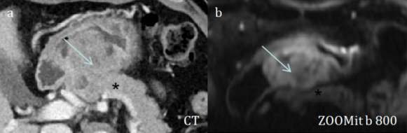
Fig. 1. CT images(a) shows that the gastric gap between the gastric antrum tumor(arrow) and the pancreatic tissue(asterisk) disappeared, indicating that the gastric cancer has invaded the pancreas. ZOOMit b=800(b) clearly shows that the contact surface of the gastric antrum tumor(arrow) and the pancreatic(asterisk) are irregular and the partial boundaries are not clear. This means that the surgical procedure was chosen for gastrojujunostomy.
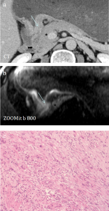
Fig.2. CT images(a) shows that the gastric gap between the gastric antrum tumor(arrow) and the pancreatic head tissue(asterisk) disappeared, indicating that the gastric cancer has invaded the pancreas. ZOOMit b=800(b) shows the contact surface of the gastric antrum tumor(arrow) with the head of the pancreas(asterisk) is smooth, indicating that radical resection can be performed. Pathology(c) confirmed tumor invasion to the muscular layer, which were arranged in duct or cord.