2564
APT combined with IVIM in differential diagnosis of endometrial carcinoma and endometrial benign lesions1Department of Radiology, the First Affiliated Hospital of Dalian Medical University, Dalian, China, 2Philips Healthcare, Beijing, China
Synopsis
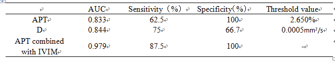
Amide proton transfer (APT) combined with intravoxel incoherent motion (IVIM) has been preliminarily applied in the diagnosis and differentiation of cervical squamous cell carcinoma, but has not been in the identification of endometrial lesions. We discussed the value of APT combined with IVIM in differentiating endometrial carcinoma from endometrial benign lesions. The AUC, sensitivity and specificity of APT combined IVIM were 0.979, 87.5% and 100%, respectively.
INTRODUCTION
Endometrial carcinoma and endometrial benign lesions have similar clinical manifestations with most of which are irregular vaginal bleeding and fluid flow, but the treatment and prognosis are quite different. Endometrial benign lesions can be treated with hysteroscopy, while endometrial carcinoma surgery still needs appropriate adjuvant treatment according to the condition. Therefore, an effective examination method to differentiate and diagnose these two diseases before operation is particularly important. As an endogenous chemical exchange saturation transfer (CEST) imaging technique, amide protron transfer (APT) imaging has received extensive attention in clinical practice [1]. Intravoxel incoherent motion (IVIM) refers to the distribution of velocity in direction or amplitude within a given voxel and measurement time, which can quantify the pure diffusion movement of water molecules in tissues and organs, measure microcirculation perfusion and other information, and comprehensively reflect the pathophysiological state of tissues [2,3]. Previous studies have shown that APT combined with IVIM technology has been preliminarily applied in the diagnosis and differentiation of cervical squamous cell carcinoma [4]. In this study, we discussed the value of APT combined with IVIM in the differential diagnosis of endometrial carcinoma and endometrial benign lesions.METHODS
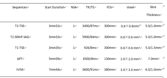
Data of 8 cases of endometrial carcinoma and 6 cases of endometrial benign lesions (5 cases of endometrial polyps and 1 case of endometrial hyperplasia) confirmed by surgery and pathology were retrospectively analyzed. All patients underwent MRI , APT and IVIM imaging on a 3.0 T MR scanner (Ingenia CX, Philips Healthcare, the Netherlands) before surgery. The scanning parameters are shown in table 1. IVIM adopted 11 b values (b= 0, 20, 50, 100, 150, 200, 400, 800, 1200, 2000, and 3000). Without knowing the pathological results, the two observers identified the lesion area of endometrium through T2WI and DWI. After the fusion of APT image and T2WI, the ROI was delineated in the corresponding lesion area, and APT value was obtained and recorded. ROI was mapped in the corresponding region of IVIM imaging to obtain the lesion's pure diffusion coefficient (D), pseudo-diffusion coefficient (D*) and perfusion fraction (f). Intra-group correlation coefficient (ICC) was used to test the consistency of measurement results between the two observers. The rank sum test was used to analyze the difference of each parameter between the two groups, and ROC was used to evaluate the diagnostic efficacy. This study has been approved by the local IRB.RESULTS
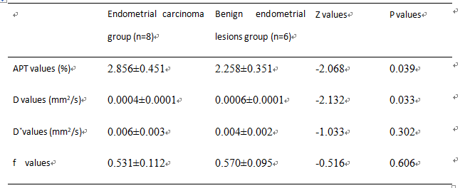
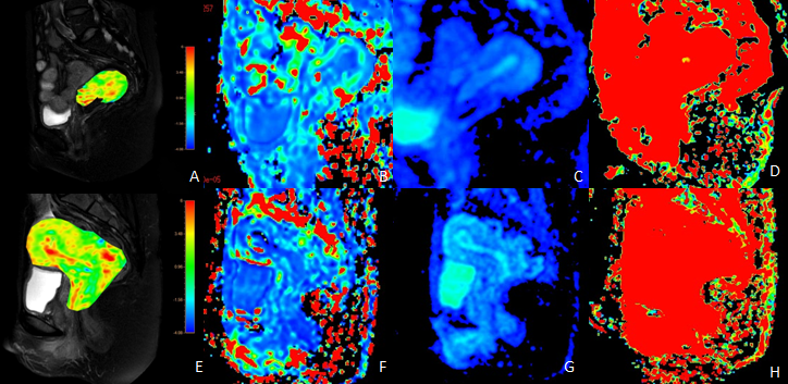
The results of parameter values and consistency test were measured by two observers in the two groups, and the numerical consistency was good in each group with ICC>0.75. APT value of endometrial carcinoma group was significantly higher than that of benign lesion group and D value of endometrial carcinoma group was significantly lower than that of benign lesion group (P<0.05). There was no statistically significant difference between the two groups in D* value and f value (P>0.05). The specific results were shown in table 2 and figure 1. The AUC values of APT, D and their combination are shown in table 3.DISCUSSION AND CONCLUSIONS
APT value in the endometrial carcinoma group was higher than that in the endometrial benign lesion group. This may due to the tumor cells in endometrial carcinoma were more metabolically active than those in the endometrial benign lesion group, and changes in protein concentration and pH value caused an increase in APT value [5]. The D value reflects the microscopic movement of water molecules. The arrangement of endometrial carcinoma cells is closer than that of endometrial benign lesions, and hence the intercellular space becomes smaller, and the movement of water molecules is limited causing the D value decreases, which is consistent with the results of this study. The AUC, sensitivity and specificity of APT combined IVIM were 0.979, 87.5% and 100%, respectively. Therefore, APT combined with IVIM can effectively differentiate endometrial carcinoma from endometrial benign lesions, which has promising clinical application value.Acknowledgements
No acknowledgement.References
[1] He YL, Li Y, Lin CY, et al. Three-dimensional turbo-spin-echo amide proton transfer-weighted MRI for cervical cancer: a preliminary study. J Magn Reson Imaging 2019. DOI: 10.1002/jmri.26710
[2] Liu J, Wan Y, Wang Z, et al. Perfusion and diffusion characteristics of endometrial malignancy based on intravoxel incoherent motion MRI at 3.0 T: comparison with normal endometrium. Acta Radiol 2016;57:1140-1148.DOI:10.1177/0284185115618550
[3] Guo L, Liu X, Liu Z, et al. Differential detection of metastatic and inflammatory lymph nodes using intravoxel incoherent motion diffusion-weighted imaging. Magn Reson Imaging 2019;65:62-66.DOI:10.1016/j.mri.2019.10.005
[4] Li B, Sun H, Zhang S, et al. The utility of APT and IVIM in the diagnosis and differentiation of squamous cell carcinoma of the cervix: A pilot study. Magn Reson Imaging 2019;63:105-113.DOI:10.1016/j.mri.2019.08.020
[5] Ray KJ, Simard MA, Larkin JR, et al. Tumor pH and Protein Concentration Contribute to the Signal of Amide Proton Transfer Magnetic Resonance Imaging. Cancer Res 2019; 79:1343-1352.DOI:10.1158/0008-5472.CAN-18-2168
Figures