2543
Improved visualization of common bile duct stone with compressed sensing MRCP1Department of Medical Imaging, Henan Provincial People’s Hospital & Zhengzhou University People’s Hospital, Zhengzhou, China, 2Siemens Shenzhen Magnetic Resonance Ltd., Shenzhen, China, 3MR Collaboration, Siemens Healthcare Ltd., Beijing, China
Synopsis
Patients with common bile duct (CBD) stone disease suffer from the relatively long scan time of magnetic resonance cholangiopancreatography (MRCP). Compressed sensing (CS) is a technique that can accelerate the speed of MRI and has been applied to MRCP. This study investigated the utility of CS-MRCP in diagnosing CBD stone disease in comparison with conventional MRCP on the 1.5T scanner. The results showed that CS-MRCP could provide comparable image quality with conventional MRCP for the diagnosis of CBD stone disease but with a substantially shorter acquisition time (1:35 vs. 4:08 mins).
Introduction
Patients with common bile duct (CBD) stone are susceptible to complications including obstructive jaundice, cholangitis, and/or acute pancreatitis1. Magnetic resonance cholangiopancreatography (MRCP) is a non-invasive diagnostic imaging modality that has been widely used for the evaluation of intra- or extrahepatic bile ducts, the pancreatic duct, and CBD stones. However, the major drawback of MRCP is the relatively long acquisition time and that it is prone to motion artifact. Compressed sensing (CS) can accelerate the speed of MR imaging in MRCP by exploiting image redundancy in sampling and reconstruction2. However, the clinical applicability of CS-MRCP in the detection of abnormalities in common bile duct stone disease has not been demonstrated on the 1.5T scanner. Therefore, the purpose of this study was to assess the utility of CS-MRCP in diagnosing CBD stone disease in comparison with conventional MRCP on the 1.5T scanner.Method
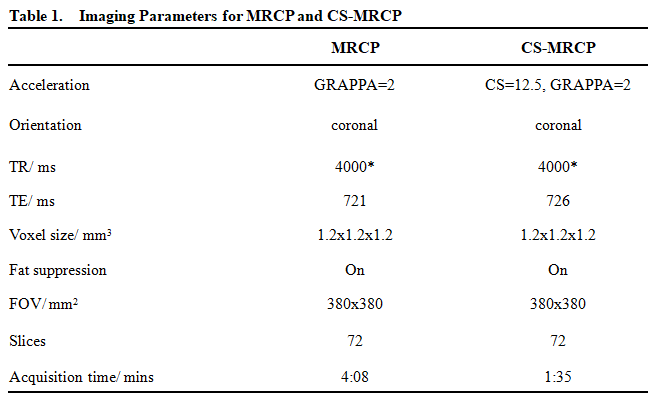
This study was approved by the institutional ethics committee. Ten patients (3 females; mean age, 57.5 years, range: 20 - 92 years) with common bile duct disease underwent CS-MRCP and conventional MRCP on a 1.5T scanner (MAGNETOM Sempra, Siemens Shenzhen Magnetic Resonance Ltd., China) with a 6-channel body phased array coil. CS-MRPCP was implemented using a prototypic compressed sensing sampling perfection with application optimized contrasts (SPACE) sequence. The acquisition parameters for these two sequences are listed in Table 1. Two radiologists independently reviewed the conventional MRCP and CS-MRCP scans and rated the image quality (IQ) for visualization of the bile duct on a 5-point Likert scale: 1 = non-diagnostic quality; 2 = poor; 3 = fair; 4 = good; and 5 = excellent. The statistical analyses were conducted using SPSS18. Wilcoxon signed rank test was used to compare the image quality scores of these two sequences. P < 0.05 was considered statistically significant.Results
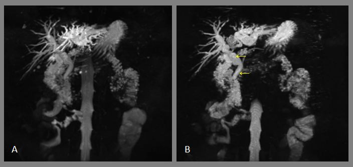
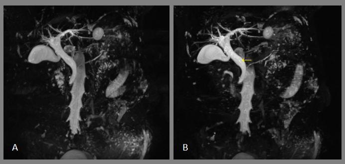
The acquisition time for CS-MRCP was reduced by more than 60% (1:35 vs. 4:08 mins) compared with the standard method in clinical practice, whereas the spatial resolution was maintained. Although there were no significant differences in the overall image quality scores between CS-MRCP and standard MRCP (mean ± standard deviation, 4.05 ± 1.25 vs. 3.85 ± 1.50, p = 0.23), the images for CS-MRCP had comparable or slightly better quality for duct and stone visualization compared with the conventional MRCP. As shown in Figures 1 and 2 that illustrated the images for two representative patients, the imaging sharpness was better, and the duct stone could be visualized more clearly in CS-MRCP.Discussion and conclusion
This study demonstrated that CS-MRCP was able to provide comparable image quality with conventional MRCP for the diagnosis of common bile duct stones but with a significantly shorter acquisition time. Although not statistically significant, the images acquired by CS-MRCP generally showed better sharpness and visualization of CBD compared with conventional MRCP, partially due to the shorter acquisition time and less motion-related imaging blurring. Furthermore, the substantially shortened scan time was particularly beneficial for patients with gallstone disease, who always suffered great pain and had a high probability of moving during the prolonged MR examination.In conclusion, CS-MRCP showed advantages over MRCP for the diagnosis of common bile duct stones and has the potential to replace MRCP in future clinical practice.
Acknowledgements
This research was supported by the National Key R&D Program of China (2017YFE0103600), National Natural Science Foundation of China (81720108021, 81601466), and Zhongyuan Thousand Talents Plan Project-- Basic Research Leader Talent (ZYQR201810117).References
1.Noda Y, Goshima S, Kojima T, et al. Improved diagnosis of common bile duct stone with single-shot balanced turbo field-echo sequence in MRCP [J]. Abdominal Radiology, 2017, 42(4): 1183-1188.
2.Taron J, Weiss J, Notohamiprodjo M, et al. Acceleration of magnetic resonance cholangiopancreatography using compressed sensing at 1.5 and 3 t: a clinical feasibility study [J]. Investigative radiology, 2018, 53(11): 681-688.
Figures