2541
Relative contribution of SWI, compared to conventional MRI, in the detection of common bile-duct (CBD) calculi1Radiology, Fortis Memorial Research Institute, Gurgaon, India, 2Clinical Science, Philips Innovation Campus, Philips India Limited, Bengaluru, Karnataka, India
Synopsis
Confident detection of small common bile duct stones is clinically challenging. Ultrasound (US) has highly variable sensitivity and the sensitivity of MRCP, while better than that of US, reduces significantly for stones<5mm. In this work, we have evaluated the relative contribution of SWI in the detection of CBD stones, relying on their susceptibility property.
Introduction
The incidence of gallstone disease stands at 15% in the USA, 22% in the Europe [1]. Out of this 5% to 20% population have choledocholithiasis in the common bile-duct (CBD) [2]. Ultrasound (US), which is clinically proven for detecting gallstone disease, has highly variable sensitivity (13% to 80%), in detecting CBD calculi [3]. Magnetic resonance cholangio-pancreatography (MRCP) is the principal technique used for the detection of gallstone disease using MR. For the detection of choledocholithiasis, while MRCP has higher sensitivity than US (81% - 93.7%), it however reduces to 64% for stones < 5mm in size[4,5]. Hence a technique for improved detection of gallstones is a mandate necessity. A recent study showed that magnetic susceptibility may be used for sensitive detection of gallbladder stones using SWI [6]. Taking cue from this work, in the current study, we evaluated the role of SWI in the detection of CBD calculi. Specifically, we evaluated the relative contribution of SWI, compared to conventional MRI techniques used in the CBD stone evaluation.Materials and Methods
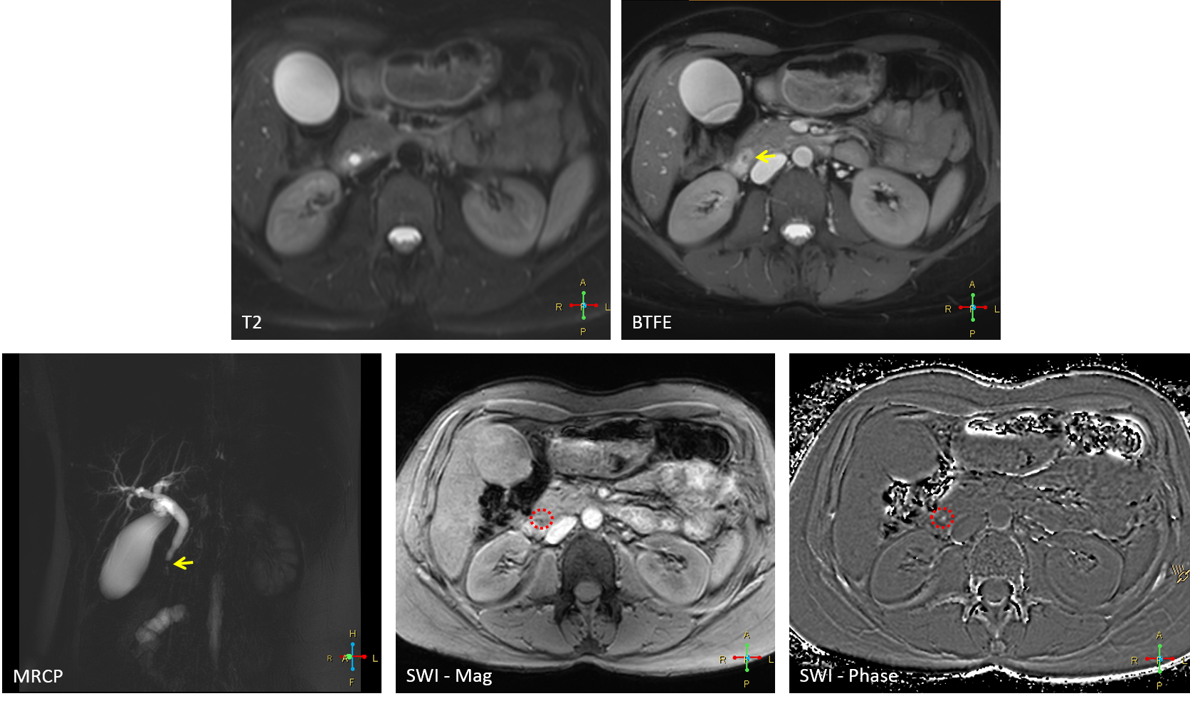
This prospective study was approved by the local institutional review committee and informed consent was obtained from the patients before MR imaging. MRI was performed on an Ingenia 3.0T (Philips, Best, Netherlands) system. SWI data acquired in patients suspected of gallstones and/or CBD stones was compared by two radiologists (with 9 and 30 years of experience in abdominal radiology) in a consensus manner. Representative MR imaging protocol parameters used are provided in Table-1 (MRCP, T2 weighted balanced turbo field echo (BTFE) and T2-weighted spin echo, SWI). Acquisition time for the 3D SWI was 17sec per breath-hold and consisted of 2 to 3 such breath-holds for covering the region of interest. All patients underwent a clinical endoscopic ultrasound (EUS) or endoscopic retrograde cholangio-pancreatography (ERCP) examination, which were used as gold standard to confirm MRI findings. The proportion of cases where calculi were visualized in SWI were noted. The relative contribution of SWI was assessed in the form of yes/no response to the following questions: findings in SWI were 1) same as conventional techniques; 2) improved diagnostic confidence; 3) was critical for diagnosis of CBD calculi. Proportion of cases falling into one of the above mentioned categories was noted for evaluating the contribution of SWI in the diagnosis of CBD stones. The size of the calculi was also evaluated wherever possible. Size of the calculi were not determined from SWI data due to confounding from the blooming effect.Results
A total of 60 patients with clinically and biochemically suspected of biliary calculi were evaluated for MRCP examination. Of these, 5 patients showed normal MRCP, 6 patients had only gallbladder stones and an additional 5 patients showed biliary sludge, which was confirmed on ERCP. Thus, N=44 cases with CBD stones, confirmed by EUS or ERCP, were reviewed as part of this study. Stones were visualized in 72.7% (32/44) of cases with MRCP, 81.2% (36/44) of cases with BTFE, 52.3% of the cases with T2 (23/44), and 100% (44) of the cases with SWI. Among these cases, SWI was found to 1) provide the same information as conventional imaging techniques in 36.6% (16/44); 2) provide improved diagnostic confidence in 31.8% (14/44); and 3) critical for diagnosis in 31.8% (14/44) of the cases. In one case, the calculi were only visualized in SWI data. Size of the stones could be evaluated in only 34 of the cases. In cases, where SWI was found ‘critical for diagnosis’, stone size could be assessed in 7 cases with an average stone size being 3.1mm ± 1.1mm. On the other hand the average stone size in the cases where they were visualized in MRCP (in 29 cases) was 6.2 ± 2.8 mm. Figure 1 shows the images from one of the cases where SWI data was critical for diagnosis.Discussion and Conclusion
This study evaluated the contribution of SWIp in the diagnosis of CBD stones, relative to the conventional MRI techniques, including MRCP and BTFE techniques. In our study cohort, while we observed that SWI had 100% sensitive in the visualization of CBD stones, we found BTFE fared slightly better than MRCP in CBD stone visualization, presumably due to T2* effects. Furthermore, our results suggest that SWI could prove critical for diagnosis in cases where stone size is small. In conclusion, we find that SWI can act as a strong adjunct to conventional MR imaging protocols used for CBD stone evaluation with very small-time penalty.Acknowledgements
No acknowledgement found.References
1. Zhang W, Jiang Z, H. T. & R, L. Epidemiology and risk factors of cholelithiasis. J Surg Concepts Pr. 16, 408–412 (2011).
2. Everhart JE, Ruhl CE. Burden of digestive diseases in the United States Part III: Liver, biliary tract, and pancreas. Gastroenterology. 2009 Apr;136(4):1134-44.
3. Cronan JJ. US diagnosis of choledocholithiasis: a reappraisal. Radiology. 1986 Oct;161(1):133-4.3.
4. Chen W, Mo JJ, Lin L, Li CQ, Zhang JF. Diagnostic value of magnetic resonance cholangiopancreatography in choledocholithiasis. World J Gastroenterol. 2015 Mar 21;21(11):3351-60.
5. Michael Maher, Jr., Adrian K. Dixon. Grainger & Allison's Diagnostic Radiology: Abdominal Imaging. 6th Edition, Elsevier Publishers. ISBN: 9780702069383
6. Gupta RK, Neelavalli J, Gupta M, Ramaniharan AK, Sinha BK, Kriplani AK, Saini J, Verma RK, Verma MK, Bhattacharjee R, Saha I. Evaluation of Gall Bladder Stones Using Susceptibility Weighted Imaging. J Comput Assist Tomogr. 2019 Sep/Oct;43(5):747-754.
Figures