2529
Use of amide proton transfer-weighted imaging to distinguish ovarian cysts due to endometriosis from cystadenoma and cystadenocarcinoma
Ye Li1, Yaxin Niu1, Xuedong Wang1, Lihua Chen1, Yanling Wu2, and Ailian Liu1
1The First Affiliated Hospital of Dalian Medical University, Dalian, China, 2Philips Healthcare, Beijing, China
1The First Affiliated Hospital of Dalian Medical University, Dalian, China, 2Philips Healthcare, Beijing, China
Synopsis
The intensity of the APT signal depends on the pH and protein content within the tissue. The later the bleeding stage is, the lower the APT value is. The ectopic endometrium repeatedly bleeds in the ovary. The blood components deposited in the cyst fluid decomposition, so the APT value of the cyst fluid is reduced. The metabolic level of tumor tissue is increased, and more protein is synthesized. Therefore, the protein content of ovarian cystadenoma and cystadenocarcinoma fluid is higher, so the APT value is increased.
Introduction
Amide proton transfer-weighted (APTw) imaging is a molecular MRI technique that generates image contrast based predominantly on the amide protons in mobile cellular proteins[1].The intensity of the APT signal depends on the pH and protein content within the tissue[2]. The study of cerebral hemorrhage[3] proved that fresh hematoma is rich in red blood cells and other plasma proteins and peptides, and the APT value is increased. The later the bleeding stage is, the lower the APT value is. The ovarian endometriotic cyst is displaced to the ovary by the endometrium. The ectopic endometrium repeatedly bleeds in the ovary. A small amount of fresh blood appears in the cystic fluid with the menstrual cycle, and the blood components deposited in the cyst fluid decomposition, so the APT value of the cyst fluid is reduced. The metabolic level of tumor tissue is increased, and more protein is synthesized. Therefore, the protein content of ovarian cystadenoma and cystadenocarcinoma fluid is higher, so the APT value is increased. Based on the above theory, we do the following research.Objective
To measure the amide proton transfer (APT) values of the cyst fluid in ovarian endometriotic cysts (OE), ovarian cystadenoma and cystadenocarcinoma(OA), and to differentiate the OE from OA.Materials and methods
A total of 15 patients with ovarian lesions who underwent pelvic MR scans at 3.0T MR scanner(Ingenia CX PHILIPS Healthcare, the Netherlands)on 2019.4-2019.10 were collected retrospectively. This research was approved by the ethics committee of the hospital. There were 4 cases of ovarian cystadenocarcinoma, 2 cases of ovarian cystadenoma, 6 cases of ovarian endometriotic cysts with 12 lesions.One case of cystic adenocarcinoma was due to smaller cystic cavity, one case of cystic adenoma and one case of ovarian endometriotic cysts (2 lesions) were due to APT without lesion level, 1 lesion of ovarian endometriotic cysts was excluded due to undistinguishable lesions (less than 1 cm in diameter).Finally, 9 lesions of OE group and 4 lesions of OA group were obtained. The scan sequence includes T1WI (axial, TR/TE:962/8ms), T2WI (axial, TR/TE:3400/87ms) and APTw (axial, TR/TE:6500/8ms, scan duration: 5min59s, Slice thinkness: 7.0mm). The APT values of the lesions were measured by two observers, three ROIs were placed on each cyst, and the mean value was calculated as the lesion APT value. The intra-class correlation coefficient (ICC) was used to test the consistency of the two observers. The ages of the two groups were compared using the Mann-Whitney U test, and the APT values of the two groups were compared using the t-test. The diagnostic value of APT values for OE was calculated using the ROC curve.Results
The two observers were in good agreement with ICC values of 0.998 and 0.967, respectively. There was no significant difference in ages between OE group (43.50±9.29) and OA group (63.75±23.50 years), p value was 0.257. The APT value of the OA group (5.20±2.19) was higher than that of the OE group (1.05±0.50), and the p value was 0.031. The APT value diagnosis OE has an AUC of 1.00, and when the APT value is ≤2.42, the diagnostic sensitivity and specificity are both 100% (Figure 1).Discussion and conclusion
The APT value of old hemorrhagic hemoglobin in the OE cyst fluid was reduced (Figure 2). The cystic fluid of OA has lots of the protein content (Figure 3), and the APT value is high. Accordingly, APT values can effectively differentiate OE from OA.Acknowledgements
No acknowledgement found.References
1.Zhou, J. , Heo, H. , Knutsson, L. , van Zijl, P. C. and Jiang, S. APT‐weighted MRI: Techniques, current neuro applications, and challenging issues. J. Magn. Reson. Imaging.2019,50: 347-364. 2. Zhou J1, Payen JF, Wilson DA, Traystman RJ, van Zijl PC. Using the amide proton signals of intracellular proteins and peptides to detect pH effects in MRI. Nat Med. 2003;9(8):1085-90.3.Ha-Kyu Jeong, Kyunghwa Han, Jinyuan Zhou, Yansong Zhao, Yoon Seong Choi, Seung-Koo Lee, Sung Soo Ahn. Characterizing Amide Proton Transfer Imaging in Hemorrhagic Brain Lesions Using 3T MRI. Eur Radiol. 2017; 27(4): 1577–1584.Figures
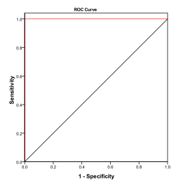
Figure 1 APT value for diagnosis of ovarian endometriosis cyst ROC curve
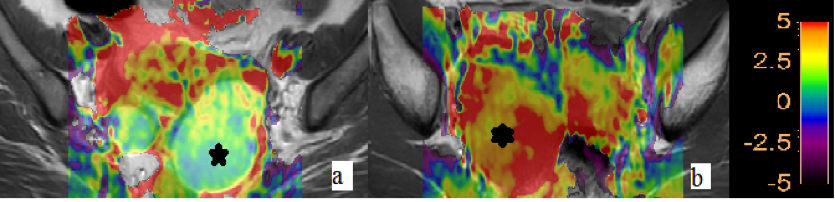
Figure 2a. Ovarian endometrial translocation cyst APT picture showed the lesion with mainly green color, indicating the APT value was 1.16. Figure 2b. Ovarian cystadenoma APT image showed the cyst mainly with red and yellow colors, indicating that the APT value was 5.5.
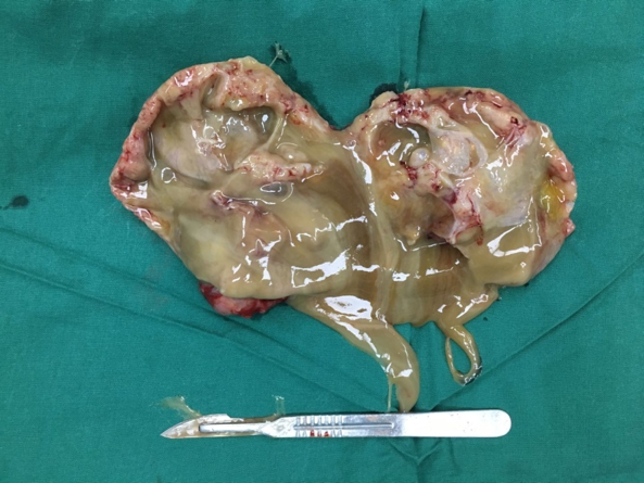
Figure 3 Intraoperative image of ovarian cystadenoma figure showed that viscous cystic fluid remained in the wall of the lesion.