2528
Application of amide proton transfer weighted imaging in differential diagnosis of endometrial carcinoma and benign endometrial lesions1First affiliated hospital of dalian medical university, Dalian, China, 2Philips Healthcare, Beijing, China
Synopsis
APT imaging has been preliminarily explored for diagnoses of cervical diseases. In this study, we investigated the potential of APTw-MRI in differential diagnosis of endometrial carcinoma and benign endometrial lesions.
INTRODUCTION
Endometrial carcinoma is a type of malignant epithelium tumor, while endometrial polyps are the most common benign endometrial lesions. It can be difficult for conventional MRI methods to distinguish these two types of endometrial lesions due to their similar signals. Amide proton transfer weighted (APTw) imaging can provide information about high-resolution distribution of protein in tumors, and thus aid in the differential diagnosis and treatment1. Previous studies have shown that the APTw-MRI can be used to study variation of APT signals for the menstrual cycle in normal uterus2, and it has also been preliminarily applied to the identification of cervical squamous carcinoma and evaluate its different grades3. In this study, we investigated the potential of APTw-MRI in differential diagnosis of endometrial carcinoma and benign endometrial lesions.METHODS
Data of 11 patients with endometrial carcinoma (group A) and 7 patients with benign endometrial lesions (group B, 5 cases of endometrial polyps and 2 cases of endometrial hyperplasia) confirmed by operation and pathology were retrospectively analyzed. All patients received MRI examinations before surgery on a 3.0 T MR scanner (Ingenia CX, Philips Healthcare, the Netherlands). The APT-weighted free-breathing 3D turbo-spin-echo sequence were scanned in the axial orientation with following scanning parameters: TR/TE = 6500/8 ms, FOV = 130 mm, voxel size = 2.0×2.0 mm2, Slice thickness = 7.0 mm, Scan duration = 5 min 59 s. APT values of both groups A and B were measured by two observers using double-blind methods. Regions of interest ROIs (diameter≥1.0cm) were placed on three adjacent layers of the largest cross section of the lesions (avoid heterogeneous areas). Means of APT values measured from the three layers were calculated and recorded. Intra-group correlation coefficient (ICC) was used to test measurement consistency between the two observers. Difference of APT values between groups A and B was analyzed by the Rank sum test, and the diagnostic efficacy was evaluated by ROC analysis.RESULTS
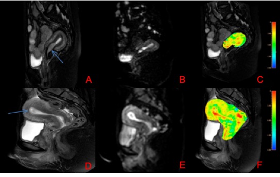
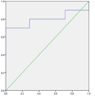
Images for two patients with endometrial carcinoma and endometrial polyp, respectively are shown in Figure 1.Measurements by the two observers are in good agreement for both group A and B (A: 0.906, B: 0.762). APT values measured for groups A (2.770 ±0.571)% were significantly higher (Z=-2.052,P<0.05=than those for group B (2.271 ±0.323)%. The AUC for APT value was 0.753 and the sensitivity was 63.6%, the specificity was 100%, with a cut-off value of 2.65 %. (Figure 2)DISCUSSION AND CONCLUSIONS
APT values of the endometrial carcinoma group were significantly higher than those of the benign endometrial lesions group. The reason may lie in that carcinoma consists of proliferative cells which produce a large amount of free protein, leading to increased APT signals, or it may because of the tumor angiogenesis that resulted in the higher APT values 4-5. In summary, the APTw-MRI may serve as an effective tool for discrimination of endometrial carcinoma from benign endometrial lesions.Acknowledgements
No acknowledgement.References
[1] He YL, Li Y, Lin CY, et al. Three-dimensional turbo-spin-echo amide proton transfer-weighted MRI for cervical cancer: a preliminary study. J Magn Reson Imaging. 2019;DOI: 10.1002/jmri.26710.
[2] Zhang S, Sun H, Li B, et al. Variation of amide proton transfer signal intensity and apparent diffusion coefficient values among phases of the menstrual cycle in the normal uterus: A preliminary study. Magn Reson Imaging. 2019;63:21-28.
[3]Meng N, Wang J, Sun J, et al. Using amide proton transfer to identify cervical squamous carcinoma/adenocarcinoma and evaluate its differentiation grade. Magn Reson Imaging. 2019;61:9-15.
[4] Ray KJ, Simard MA, Larkin JR, et al. Tumor pH and protein concentration contribute to the signal of amide proton transfer magnetic resonance imaging. Cancer Res. 2019; 79:1343-1352.
[5] Li B, Sun H, Zhang S, et al. The utility of APT and IVIM in the diagnosis and differentiation of squamous cell carcinoma of the cervix: A pilot study. Magn Reson Imaging. 2019;63:105-113.
Figures