2525
Application value of DKI combined with APT in differentiating pathological grades of squamous cell carcinoma of cervix1The First Affiliated Hospital of Dalian Medical University, Dalian Medical University, Da Lian, China, 2Philips Healthcare, Beijing, China, Bei jing, China
Synopsis
We aimed to explore the value of amide proton transfer-weighted (APTw) imaging combined with diffusion kurtosis imaging (DKI) in differentiating pathological grades of squamous cell carcinoma (SCC) of cervix. The result showed that the highest diagnostic efficacy (AUC: 0.971) was acquired using APTw combined with mean kurtosis (MK).
Introduction
Squamous cell carcinoma (SCC) is the most common pathological type of cervical carcinoma, the degree of pathological grading affects the treatment and prognosis [1]. Preoperative staging is judged by gynecological examination, which is highly subjective. MR has gradually become an imaging technology for diagnosis of pelvic lesions due to the high soft tissue resolution [2]. Amide proton transfer-weighted (APTw) imaging technology can provide the information of high revolution free protein and amino compound proton of peptide in vivo to reflect the distribution of protein in the tumor and aid in tumors grading and staging. Diffusion kurtosis imaging (DKI) is more sensitive to the heterogeneity of water molecule diffusion, so it has the ability to reflect the microstructure information of tissue more accurately [3, 4]. In this study, we demonstrated that APTw combined with DKI could improve the diagnostic efficiency.Purpose
To explore the value of quantitative parameters of DKI combined with APTw in differentiating pathological grades of squamous cell carcinoma (SCC) of cervix.Materials and Methods
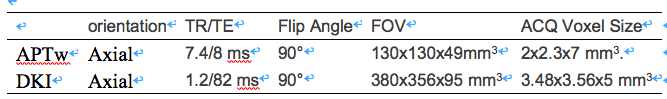
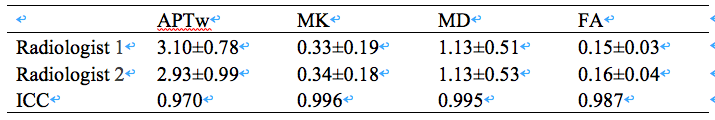
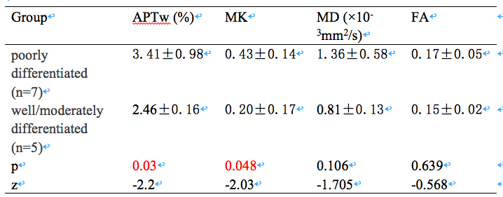
Twelve patients with cervical SCC (age 52.42 ± 7.63) underwent MR examination for DKI and APTw on 3.0T MR (Ingenia CX, Philips). According to the pathological results, they were divided into poorly differentiated(7 patients) and well/moderately differentiated(5 patients) groups. APTw images were transferred to a workstation (Intellispace Portal, Philips Healthcare) for post-processing. The scan parameters were shown in Table 1. DKI raw data was transferred to GE AW 4.6 workstation and the DKI parameter maps were calculated in Functool software. The mean kurtosis (MK), mean diffusion (MD), fractional anisotropy (FA) and APTw values of the SCC were measured by two radiologists (Fig 1). The DKI parameters and APTwvalue between two groups were compared. The diagnostic efficacy of DKI combined with APT for differentiating pathological grades was analyzed by logistic regression. Intra-group correlation coefficient (ICC) was used to test the consistency of the two observers' measurements. The difference in parameters was analyzed by Mann-WhitneyU test, and the diagnostic efficacy was evaluated by ROC analysis.Results
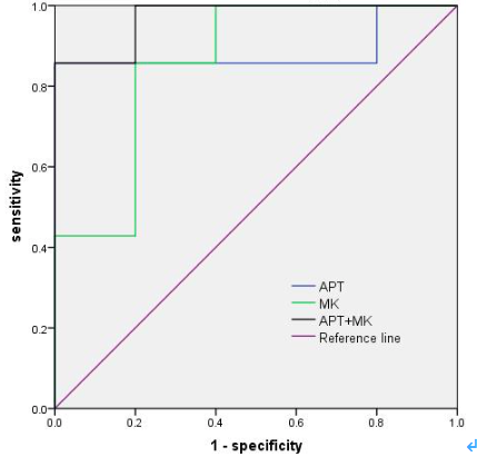
The consistency of the measurements obtained by the two observers were good (ICC value > 0.8, Table 2). The value of APTw, MK, MD, and FA in poorly differentiated group was higher than that of well/moderately differentiated group, with a statistically significant difference (P values of APTw and MK < 0.05, while P values of MD and FA > 0.05, Table 3). Area under the curve (AUC) for APTw and MK to differentiate poorly differentiated form well/moderately differentiated SCC were 0.886 and 0.857. The sensitivity of above parameters corresponding to their respective feasible threshold values were 85.7% and 100%, and the specificity were 85.7% and 80%. With APTw values combined with MK in differentiating pathological grades of SCC, we found that the diagnostic efficacy was significantly higher (AUC=0.971, Fig 2).Discussion and conclusion
APTw imaging combined with DKI has a high value in differentiating pathological grades of SCC. Among the single parameter, the diagnostic efficacy of APTw was the highest (AUC: 0.886). When APTw values combined with MK, the diagnostic efficacy was significantly improved (AUC: 0.971). APTw imaging can contribute to the differentiating pathological grades of SCC which possess different water-exchangeable chemical groups and tissue physicochemical properties. Compared with well/moderately differentiated SCC, the diffusion in poorly differentiated SCC was limited, so MK value was larger [5].Acknowledgements
No acknowledgement foundReferences
[1] Pedro T. Ramirez,Rene Pareja,Gabriel J. Rendón,Carlos Millan,Michael Frumovitz,Kathleen M. Schmeler. Management of low-risk early-stage cervical cancer: Should conization, simple trachelectomy, or simple hysterectomy replace radical surgery as the new standard of care? [J]. Gynecologic Oncology,2014,132(1).[2] Daido s, Kido A, Kataoka M, et al. MR imaging of uterine morphology and dynamic changes during lactation[J]. J Magn Reson Imaging. 2017 Feb;45(2):617-623.
[3] Wu G, Li MM, Chen F, et al.Diffusion-kurtosis imaging predicts early radiotherapy response in nasopharyngeal carcinoma patients.Oncotarget, 2017, 8:66128-66136.
[4] He Yong-Lan, Li Yuan, Lin Cheng-Yu et al. Three-dimensional turbo-spin-echo amide proton transfer-weighted mri for cervical cancer: A preliminary study. [J] Magn Reson Imaging, 2019, 50: 1318-1325.
[5]Pavilla A, Gambarota G, Arrigo A, et al.Diffusional kurtosis imaging (DKI) incorporation into an into an intravoxel incoherent motion (IVIM) MR model to measure cerebral hypoperfusion induced by hyperventilation challenge in healthy subject. MAGMA, 2017, 30:545-554.
Figures