2523
Compressed Sensing with and without Deep Learning Reconstruction: Comparison of the Utility for Women’s Pelvic MRI with Parallel Imaging1Radiology, Fujita Health University School of Medicine, Toyoake, Japan, 2Canon Medical Systems Corporation, Otawara, Japan, 3Radiology, Fujita Health University Hospital, Toyoake, Japan, 4Joint Research Laboratory of Advanced Medical Imaging, Fujita Health University School of Medicine, Toyoake, Japan
Synopsis
There have been no major reports for assessing the utility of compressed sensing (CS) and deep learning reconstruction (DLR) on women’s pelvic MRI as compared with routinely applied parallel imaging (PI). We hypothesized that CS with DLR was able to improve image quality and shorten examination time on women’s pelvic MRI, when compared with PI. The purpose of this study was to directly compare the utility of CS and DLR with PI at women’s pelvic MRI examination in patients with different women’s pelvic diseases.
Introduction
Since the beginning of 2000s, improving temporal and spatial resolution on women’s pelvic MR imaging (MRI) have been tried to using not only image domain based parallel imaging (PI) techniques, but also k-space domain based parallel imaging technique at 1.5 and 3 Tesla (T) MR systems (1). However, the reduction of examination time, temporal and spatial resolution by PI have been suggested as limited due to increasing the number of coil element. Recently, compressed sensing (CS) is suggested as a new method for aiming to reduce the number of k-space samples by exploiting compressibility or sparsity in an appropriate transform domain (2). However, one of the drawbacks of CS is suggested as relatively lower SNR than PI. In this situation, deep learning reconstruction (DLR) was also introduced for improving imaging quality of not only central nervous system, but also body MRIs (3). However, there have been no major reports for assessing the utility of CS and DLR on women’s pelvic MRI as compared with routinely applied PI. We hypothesized that CS with DLR was able to improve image quality and shorten examination time on women’s pelvic MRI, when compared with PI. The purpose of this study was to directly compare the utility of CS and DLR with PI at women’s pelvic MRI examination in patients with different women’s pelvic diseases.Materials and methods
Thirty consecutive women’s pelvic disease patients (mean age 50 years, range 26–82 years) underwent conventional T2-weighted imaging at 3T MR system (Vantage Galan 3T, Canon Medical Systems, Otawara, Japan) by PI (SPEEDER, Canon Medical) and CS (Compressed SPEEDER, Canon Medical). Acceleration factors of PI and CS were 1.5 and 3 in this study. Then, all CS images were reconstructed with and without DLR. As quantitative image quality indexes in each patient, SNR, percentage of coefficient of variation (%CV) and contrast–to–noise ratio (CNR) were determined by ROI measurement. For qualitative image quality assessment, two board certified radiologists evaluated overall image quality, artifacts and diagnostic confidence by 5–point scoring system, and final value of each qualitative index was made by consensus of two readers. To compare quantitative image quality indexes among three methods, SNR, %CV and CNR were compared among PI and CS with and without DLR by Wilcoxon signed-rank test. For evaluating inter-observer agreement of each qualitative index, weighted kappa statistics and c2 test was performed. To compare each qualitative index among three methods, each index was also compared among PI and CS with and without DLR by Wilcoxon signed-rank test. Finally, mean examination time was also compared among PI and CS with and without DLR by Tukey’s HSD test .Results
Representative case is shown in Figure1. Figure 2 shows the compared result of each quantitative image quality index among three methods. When compared SNR, SNR of CS with DLR were significantly higher than that of others (p<0.05). Moreover, SNR of PI was significantly higher than that of CS without DLR (p<0.05). On comparison of %CV, %CV of CS with DLR was significantly smaller than that of others (p<0.05). In addition, %CV of PI was also smaller than that of CS without DLR (p<0.05). When compared CNR, CNR of PI and CS with DLR were significantly higher than that of CS without DLR (p<0.05), although there was no significant difference of CNR between PI and CS with DLR. Figure 3 shows the results of inter-observer agreement for each qualitative image quality index between two readers. On visually assessed each qualitative image quality, inter-observer agreements for overall image quality and artifact of PI and CS without DLR were assessed as almost perfect (0.93≤κ≤0.95, p<0.0001), although those of CS with DLR were determined as substantial or moderate (overall image quality: κ=0.72, p<0.0001; artifacts: κ=0.59, p<0.0001). On the other hand, inter-observer agreements for diagnostic confidence on all methods were assessed as equal and substantial (κ=0.65, p<0.0001). Figure 4 shows the results of comparison for each qualitative image quality index among three methods. Overall image quality scores of PI and CS with DLR were significantly higher than that of CS without DLR (p<0.0001). Moreover, artifact scores of PI and CS with DLR were significantly lower than that of CS without DLR (p<0.0001). On the other hand, there were no significant differences of diagnostic confidence among PI and CS with and without DLR (p>0.05). Mean examination times of CS with and without DLR (with DLR: 119.9±15.0s, without DLR: 112.0 ± 14.0s ) were significantly shorter than that of PI (214 .1± 26.1s, p <0.0001).Conclusion
CS is able to significantly reduce examination time than PI, and DLR can significantly improve quantitative and qualitative image qualities of MR imaging obtained with CS. When compared with PI, combination of CS and DLR makes it possible to improve women’s pelvic MR examination in patients with different diseases.Acknowledgements
No acknowledgement found.References
- Deshmane A, et al. J Magn Reson Imaging. 2012 Jul;36(1):55-72.
- Feng L, et al. J Magn Reson Imaging. 2017 Apr;45(4):966-987.
- Kidoh M, et al. Magn Reson Med Sci. 2019 Sep 4. doi: 10.2463/mrms.mp.2019-0018. [Epub ahead of print]
Figures
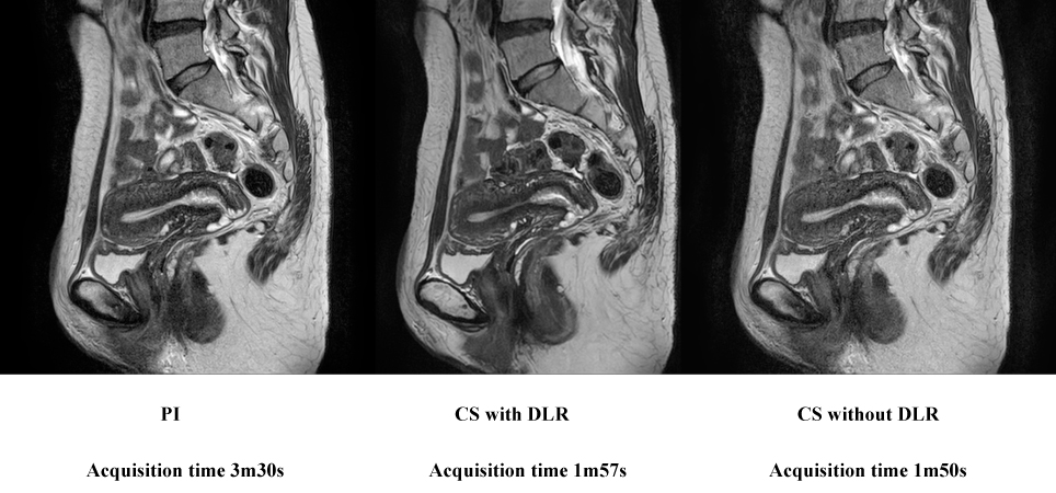
Figure 1. 43–year old female patient with Nabothian cyst (L to R: PI, CS with DLR and CS without DLR).
All images clearly demonstrate Nabothian cyst as well as normal structures in not only uterus, but also others. On quantitatively and qualitatively assessed indexes, CS with DLR was higher than that of others. In addition, these indexes of CS without DLR were close to those of PI.
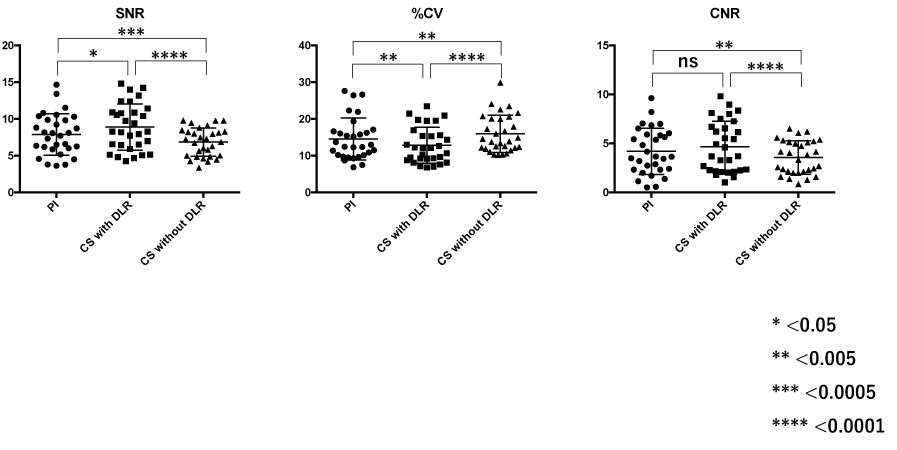
Figure 2. Results of comparison for each quantitative image quality index among three methods.
SNR and %CV of CS with DLR were significantly superior to those of others (p<0.05). However, there was significant difference between PI and CS without DLR (p<0.05). On the other hand, CNR of PI and CS with DLR were significantly superior to that of CS without DLR (p<0.05).
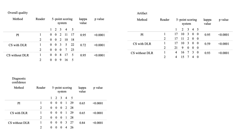
Figure 3. Results of inter-observer agreement for each image quality index on three methods.
Inter-observer agreements for overall image quality and artifact of PI and CS without DLR on three methods were assessed as follows: PI, 0.65≤κ≤0.95; CS with DLR, 0.59≤κ≤0.72; CS without DLR, 0.84≤κ≤0.95. almost perfect (0.93≤κ≤0.95), although those of CS with DLR were determined as substantial or moderate (overall image quality: κ=0.72, artifacts: κ=0.59). On the other hand, inter-observer agreements for diagnostic performance on all methods were assessed as substantial (κ=0.65)
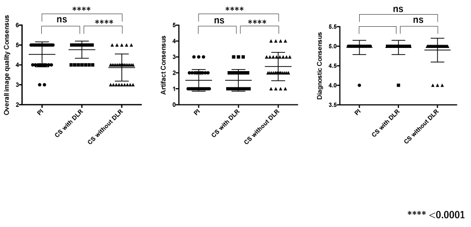
Figure 4. Results of comparison for each qualitative image quality index among three methods.
Overall image quality and artifact scores of PI and CS with DLR were significantly better than those of CS without DLR (p<0.0001), although there was no significant difference among PI and CS with and without DLR.