2520
Effect of different Compressed-SENSE acceleration factors on pancreas volume using 3D mDIXON Quant1The first affiliated hospital of Dalian Medical University, Dalian, China, 2Philips Healthcare, Beijing, China
Synopsis
The pancreatic volume can reflect the function of the pancreas to a certain extent. The 3D mDixon Quant can be used to assess the volume of the tissue structure, but some patients cannot tolerate the long-term breath test. This study was designed to ensure pancreas volume using different CS acceleration factors on the premise of ensuring image quality. The results show that CS-SENSE 6 guarantees image quality and reduces scan time.
Introduction
The pancreas plays a central role in metabolism and is involved in the pathogenesis of several diseases. Pancreas volume is a holistic quantitative measure of pancreas size but measurements of pancreas size and composition are usually time-consuming and operator-dependent1.Diabetes can also lead to a corresponding change in pancreatic volume 2. Therefore, the study of pancreatic volume has important clinical significance for the prevention, diagnosis, treatment and prognosis evaluation of related diseases. However, most pancreatic volume assessment is limited to computed tomography (CT) studies requiring radioactive agents3. 3D mDIXON Quant was found to enable robust water-fat separation and may be used in pancreatic volume. The purpose of this study was to evaluate the feasibility of pancreatic volume measurement using 3D mDIXON Quant technique and the effect of different Compressed SENSE acceleration factors on the premise of ensuring image quality.Materials and method
Institutional review board approval and informed consent were obtained. 10 healthy volunteers (4 males and 6 females, mean age 24.91±1.64 years, age range 22-27years, BMI range 17.71-28.73kg/m2, mean BMI 21.75±3.35 kg/m2) were scheduled for pancreas 3D mDIXON MR imaging on a 3T MR scanner (Ingenia 3.0T CX; Philips Healthcare, Best, the Netherlands). Scan parameters were as follows: FOV=375mm×300mm, TR/TE=6ms/XXms, Slice thickness and gap=5.0mm/2.5mm, SENSE =2, 4, Compressed-SENSE(CS)=2, 4, 5, 6, Echo=6. Data was transferred to the IntelliSpace Portal, (Philips Healthcare). Pancreatic volume was measured using one single-layer ROI drawing superposition volume technology as shown in Figure.1. ROIs (100mm2) were placed on the maximal level of the head, body, tail, and bilateral erector spinae of each sequence, and the signal value and noise were measured (Fig.2). The measured pancreatic and erector spinae data were averaged, and the SNR and CNR were calculated. The Friedman test was used to compare the SD value, SNR, CNR, and pancreatic volume between the sequences. P < 0.05 is considered to be statistically significant. This study has been approved by the local IRB.Results
There was no significant difference in SD value, SNR, CNR between groups(P>0.05). The pancreatic volume (cm3) of SENSE 2, 4, CS-SENSE 2, 4, 5, 6 are 61.3±8.5, 59.5±8.4, 59.3±7.6, 58.9±9.8, 59.5±8.7, 61.3±7.8. There was no significant difference in the volume among the groups (P > 0.05) (Table1).Discussion
The 3D mDIXON Quant sequence scans 90 layers, collects 6 echoes in one breath, analyzes 7 fat peaks, and combines T2* correction to generate FF maps and T2* maps. The quantitative pancreatic volume obtained in this study is comparable with the results of former studies in Asian1.The CS technology used in our research uses digital random sparse sampling to ensure the fidelity of the image. The signal acquired in K space is converted to Hilbert space via Fourier transform and wavelet transform (H space) 4. Using CS 6 can significantly reduce the scan time while ensuring image quality.Conclusion
3D mDIXON Quant technique can be used for pancreatic volume quantitative analysis. Compressed SENSE technique can be used for the acquisition acceleration in the 3D mDIXON Quant data acquisition. Compressed SENSE acceleration factor 6 can shorten the scanning time with the guarantee of the image quality.Acknowledgements
No acknowledgement found.References
- Steve V. DeSouza, Ruma G. Singh, Harry D. Yoon, Rinki Murphy, Lindsay D. Plank & Maxim S. Petrov (2018): Pancreas volume in health and disease: a systematic review and meta-analysis, Expert Review of Gastroenterology & Hepatology, DOI: 10.1080/17474124.2018.1496015
- Sepe Paul S,Ohri Ashray,Sanaka Sirish et al. A prospective evaluation of fatty pancreas by using EUS[J]. Gastrointest. Endosc., 2011, 73(5): 987-93.
- Saisho Y, Butler AE, Meier JJ, Monchamp T, Allen-Auerbach M, et al... (2007) Pancreas volumes in humans from birth to age one hundred taking into account sex, obesity, and presence of type-2 diabetes. Clin Anat 20: 933–942.
- Hannukainen Jarna C,Borra Ronald,Linderborg Kaisa et al. Liver and pancreatic fat content and metabolism in healthy monozygotic twins with discordant physical activity[J]. J. Hepatol., 2011, 54(3): 545-52.
Figures
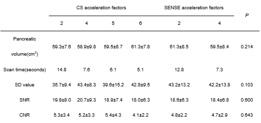
Table 1 Differences in pancreas volume, scan time, CNR, SNR of different CS and SENSE acceleration factors
There was no significant difference in SD value, SNR, CNR between groups(P<0.05). There was no significant difference in the volume among the groups(P>0.05).
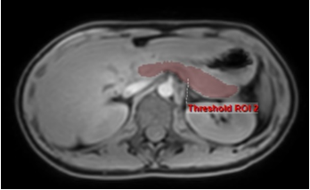
Fig.1 3D mDIXON Quant water phase
Female, 27 years old, BMI17.71kg/m2. Volume of whole pancreas is extracted. The pancreatic volume was automatically calculated 67.73cm3.
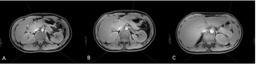
Fig.2 3D mDIXON Quant water phase
ROIs were placed on the maximal level of the head(A), body(B), tail(C), and bilateral erector spinae of each sequence, and the signal value and noise were measured.