2499
Comparison of T1rho and Diffusion Kurtosis Imaging in Evaluation of Hepatic Sinusoidal Obstruction Syndrome in Rats1School of Biomedical Engineering, Southern Medical University, Guangzhou, China, 2Guangdong Provincial Key Laboratory of Medical Image Processing, Southern Medical University, Guangzhou, China, 3Department of Medical Imaging Center, Nanfang Hospital, Southern Medical University, Guangzhou, China, 4Key Laboratory of Mental Health of the Ministry of Education, Southern Medical University, Guangzhou, China, 5Philips Healthcare, Guangzhou, China, 6Department of MRI, The First People’s Hospital of Foshan (Affiliated Foshan Hospital of Sun Yat-sen University), Foshan, China
Synopsis
T1rho represents the spin-lattice relaxation time constant in the rotating frame, which sever as a biomarker for liver function associated with alteration in the macromolecular content of tissues. Diffusion kurtosis imaging (DKI) was developed to measure non-Gaussian diffusion, which have increasingly been used to characterize microstructural heterogeneity in vivo. Sinusoidal obstruction syndrome (SOS) is a dynamic process with complex histopathological changes in liver. Our study attempts to investigate the relationship between T1rho and DKI parameters in the light of pathological examinations to help us better understand the contribution of the possible factors to changes in T1rho.
Synopsis
T1rho represents the spin-lattice relaxation time constant in the rotating frame, which sever as a biomarker for liver function associated with alteration in the macromolecular content of tissues. Diffusion kurtosis imaging (DKI) was developed to measure non-Gaussian diffusion, which have increasingly been used to characterize microstructural heterogeneity in vivo. Sinusoidal obstruction syndrome (SOS) is a dynamic process with complex histopathological changes in liver. Our study attempts to investigate the relationship between T1rho and DKI parameters in the light of pathological examinations to help us better understand the contribution of the possible factors to changes in T1rho.Introduction
Hepatic sinusoidal obstruction syndrome is a dynamic liver injury accompanied by sinusoidal deformity, necrosis, inflammation, and fibrosis in the process, ultimately resulting in liver dysfunction1. Diffusion kurtosis imaging (DKI) is based on the non-Gaussian diffusion model, which can not only detect restricted water diffusion (Dapp), but also characterize the heterogeneity of tissue microstructure (Kapp) 2. T1 relaxation time in the rotating frame (T1rho), which was sensitive to slow motion interactions between motion-restricted water molecules and the local macromolecular of tissue, was reported as a feasible biomarker for the assessment of liver function and fibrosis 3,4. In this study, we attempted to identify possible influencing factors of T1rho in the liver with SOS using DKI and pathological examination as references.Methods
Study design: Nine Sprague-Dawley rats weighing from 220–250g were included. Six rats were gavaged with 10mg/ML monocrotaline (MCT) solution at a dose of 160mg/Kg body weight, another three rats were treated as baseline without any intervention. Follow-up imaging was performed at three timepoints: baseline rats underwent MRI scan on 1 day before gavage, three MCT intragastrical rats were selected for MRI scanning on 3 and 7 days after gavage, respectively. The rats were sacrificed for histological examination immediately after MRI scan.MRI: Imaging was carried out on a 3.0 T MRI scanner (Philips Healthcare, Netherlands). Diffusion-weighted MRI was performed with a single-shot spin-echo echo-planar imaging (EPI) sequence. The parameters were as follows: TR/ TE = 2000/68 ms, EPI factor = 59, FOV=60×60 mm, slice thickness = 3 mm, number of slices = 13, matrix = 64×64. Five b values were selected: 0, 500, 1000, 1500, 2000 s/mm2 and apply in three diffusion directions.
Image analysis: T1rho and DKI data was postprocessed using a manufacturer supplied software and performed pixel-by-pixel fitting. Five ROIs were manually drawn by a radiologist on the largest cross-section of liver (Figure 1). The mean measured for the five ROIs was treated as the parameter value.
Histology: For pathological analysis, liver samples were stained with hematoxylin and eosin, and Masson trichrome. According to the previous study 1, seven pathological features were scored with 4-point criteria (0, absent; 1, mild; 2, moderate; and 3, severe), The pathological score was calculated by adding up the individual scores.
Results
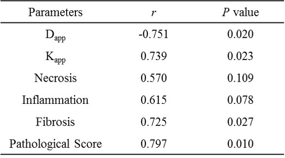
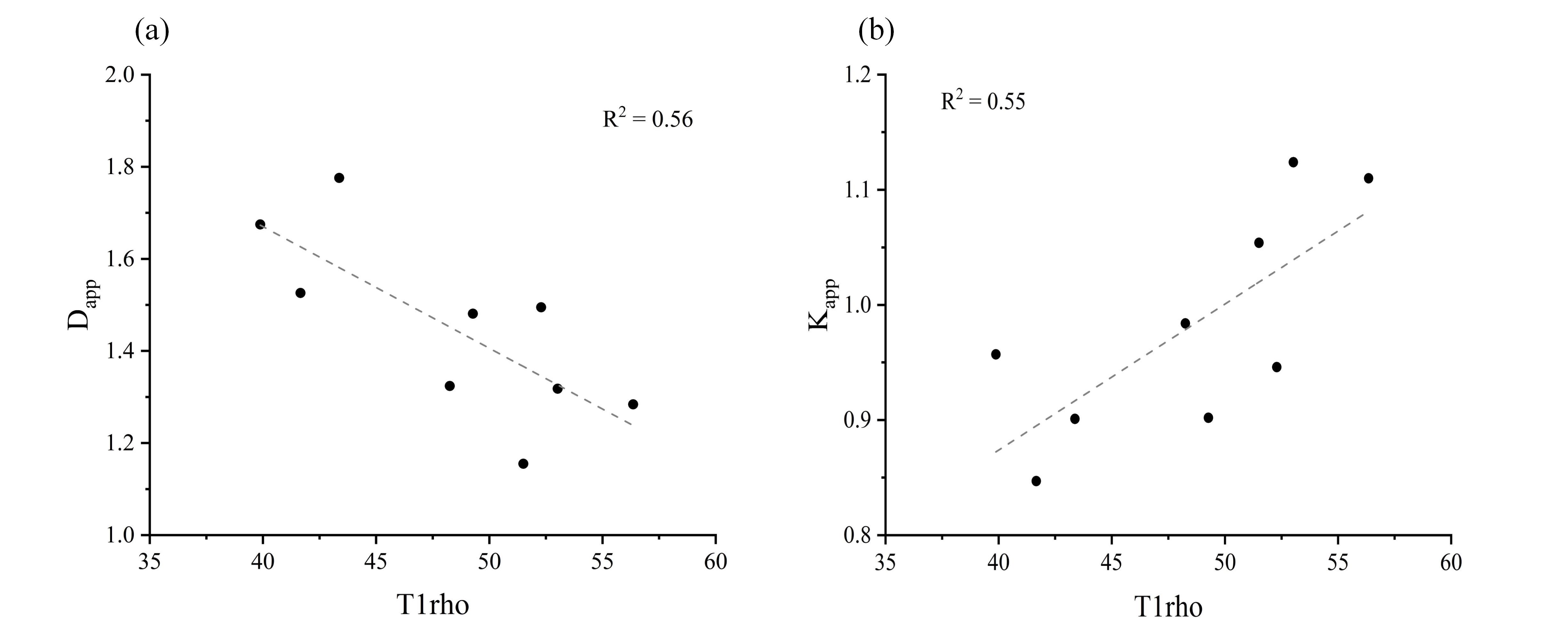
As shown in Figure 2, T1rho and Kapp were positively correlated with the pathological score, necrosis, inflammation and fibrosis. In addition, T1rho was negatively correlated with Dapp. Although T1rho was significantly correlated with fibrosis in comparisons with individual pathological feature, it was not significantly correlated with necrosis and inflammation (Table 1). The T1rho values showed strong correlations with Dapp and Kapp derived from DKI (Figure 3).Discussion
In this study, we have shown that T1rho was significantly correlated with liver injury in SOS progression. Strong correlation between T1rho and DKI metrics indicated that the change of T1rho value could be affected by multiple pathological features. The fundamental principle resulted in elevated T1rho in the liver has still not been fully investigated, and it remains unclear what factors contribute to T1rho relaxation. Our results demonstrated that T1rho was more sensitive with fibrosis than necrosis and inflammation. However, the increment of T1rho between baseline and day 3 may not result from the fibrosis, since only one liver sample showed a small amount of fibrosis. During early SOS progression, there is an increase in extracellular matrix, composed of accumulation of macromolecules and cellular matrices. In this regard, we speculated that T1rho relaxation might be affected by the accumulation of fibril-forming collagen or the decrease of mobile water molecule in SOS deterioration.Conclusion
T1rho was a feasible tool in detection of SOS progression. Although the specific factor leading to the increase of T1rho value in SOS detection have not been clearly explained, we have reason to believe that there are multiple contributing factors for T1rho increment in hepatic SOS. T1rho relaxation was a complex process involving a combination of biological, chemical and physical factors, further study will be necessary to clarify these contributions and make T1rho as a reliable imaging biomarker.Acknowledgements
No acknowledgement found.References
1. DeLeve LD, McCuskey RS, Wang X, et al. Characterization of a reproducible rat model of hepatic veno-occlusive disease. Hepatology 1999;29(6):1779-1791.
2. Jensen JH, Helpern JA, Ramani A, Lu H, Kaczynski K. Diffusional kurtosis imaging: the quantification of non-gaussian water diffusion by means of magnetic resonance imaging. Magn Reson Med 2005;53(6):1432-1440.
3. Allkemper T, Sagmeister F, Cicinnati V, et al. Evaluation of fibrotic liver disease with whole-liver T1rho MR imaging: a feasibility study at 1.5 T. Radiology 2014;271(2):408-415.
4. Wang YX, Yuan J, Chu ES, et al. T1rho MR imaging is sensitive to evaluate liver fibrosis: an experimental study in a rat biliary duct ligation model. Radiology 2011;259(3):712-719.
Figures