2486
Radiomics Analysis on Gd-EOB-DTPA-Enhanced MRI for Prediction of Liver Function and Hepatic Cirrhosis1Radiology, Central Hospital of Wuhan, Tongji Medical College, Huazhong University of Science and Technology, Wuhan, China, 2Clinical Science,Philips Healthcare, Shanghai, China
Synopsis
This retrospective study explored the value of a radiomics-based model on Gd-EOB-DTPA-Enhanced MRI for predicting liver function and cirrhosis in clinic. Multi-class radiomics feature extraction was performed on 2D-view whole liver at portal level on HBP MRI obtained 20 min after Gd-EOB-DTPA-enhanced MRI. A prediction model including 15 radiomics features using a machine learning logistic regression classifier showed the mean AUCs on train dataset and test dataset were 0.91 and 0.87 for diagnosing Child-Pugh A respectively; 0.93 and 0.93 for diagnosing liver cirrhosis, respectively.
Synopsis
This retrospective study explored the value of a radiomics-based model on Gd-EOB-DTPA-Enhanced MRI for predicting liver function and cirrhosis in clinic. Multi-class radiomics feature extraction was performed on 2D-view whole liver at portal level on HBP MRI obtained 20 min after Gd-EOB-DTPA-enhanced MRI. A prediction model including 15 radiomics features using a machine learning logistic regression classifier showed the mean AUCs on train dataset and test dataset were 0.91 and 0.87 for diagnosing Child-Pugh A respectively; 0.93 and 0.93 for diagnosing liver cirrhosis, respectively. Radiomics analysis of gadoxetic acid-enhanced HBP images allows for accurate diagnosis of clinically significant liver function reservation and liver cirrhosis and may be a promising noninvasive method for assessment of liver cirrhosis.Introduction
Liver fibrosis is an important cause of morbidity and mortality in patients with chronic liver disease, and complications mainly occur in patients with advanced fibrosis (1). Although liver biopsy is the current reference method for staging liver fibrosis, it has limitations, including risks of procedure-related complications, sampling error, and inter-observer variability (2). Thus, there is a need for simple, noninvasive, and accurate methods to diagnose and stage liver fibrosis. Gd-EOB-DTPA (Primovist; Bayer Health Care, Berlin, Germany), a liver specific contrast agent, has shown a robust value for diagnosis of focal hepatic lesions, and may allow for the assessment of liver fibrosis (3)because the hepatic uptake of gadoxetic acid depends on liver function, and the degree of liver enhancement on hepatobiliary phase (HBP) images reflects the degree of liver fibrosis (4). Moreover, enhancement with gadoxetic acid may help radiologists to view fibrosis-associated changes in liver texture by improving contrast between enhanced hepatocytes and unenhanced fibrotic scars. Previous studies have shown the value of gadoxetic acid–enhanced MRI in assessing liver cirrhosis evaluated by visual assessment, the degree of liver enhancement, and histogram analysis of liver signal heterogeneity on HBP images(5). However, these studies relied on visual assessment or a single quantitative parameter for assessing liver fibrosis, which may not be sufficient for full and objective evaluation of imaging features. Radiomics analysis is a mathematical statistical procedure to extract objective and quantitative parameters (signal intensity, histogram-based features, and texture features) from given images. Here, we report a model based on radiomics features extracted from gadoxetic acid–enhanced HBP images for evaluating liver function and hepatic cirrhosis. In this retrospective study, we used radiomics analysis of 2D-view whole liver at portal level on HBP MRI to investigate the values of prediction of Child-Pugh A and hepatic cirrhosis.Purpose
To develop and validate a radiomics-based model for evaluating liver function and hepatic cirrhosis by using Gd-EOB-DTPA–enhanced hepatobiliary phase MRI.Methods
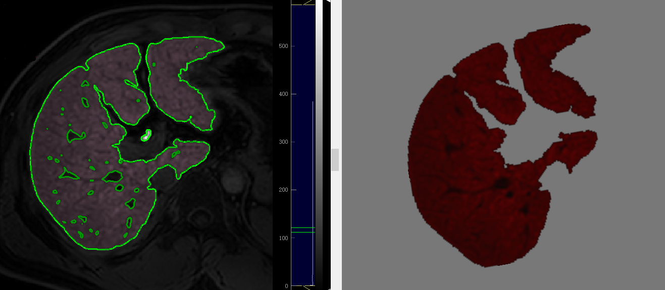
In this retrospective study, 214 patients with pathologic analysis-proven liver fibrosis who underwent gadoxetic acid–enhanced MRI from June 2017 to September 2019 were randomized into training and testing cohorts at a ratio of 7:3, respectively. For each patient, 1227 radiomics features were calculated from Gd-EOB-DTPA–enhanced hepatobiliary phase MRI using pyradiomics. In the training dataset, we normalized all the features using Min-Max scaling algorithm. The Spearman correlation between each radiomics feature and the classification label was calculated and features with the coefficient lower than 0.2 or the corresponding p-value greater than 0.05 were removed accordingly. To do the further dimensionality reduction, least absolute shrinkage and selection operator (LASSO) algorithm was applied and the remaining features were used to train a prediction model. We used 5-fold cross-validation to evaluate the performance of a specific classifier. For all the two classifications, Logistic Regression was found to produce the most accurate model in training dataset. The performance of the trained models was further evaluated in the independent testing datasets, respectively.Results
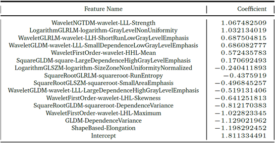
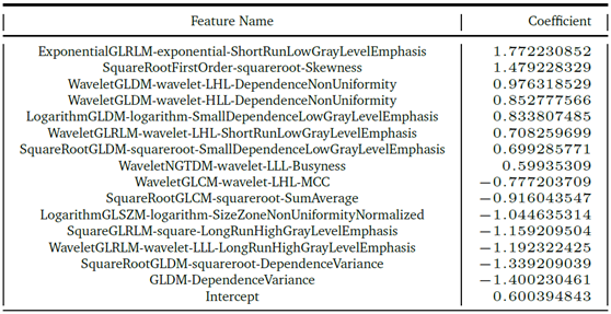
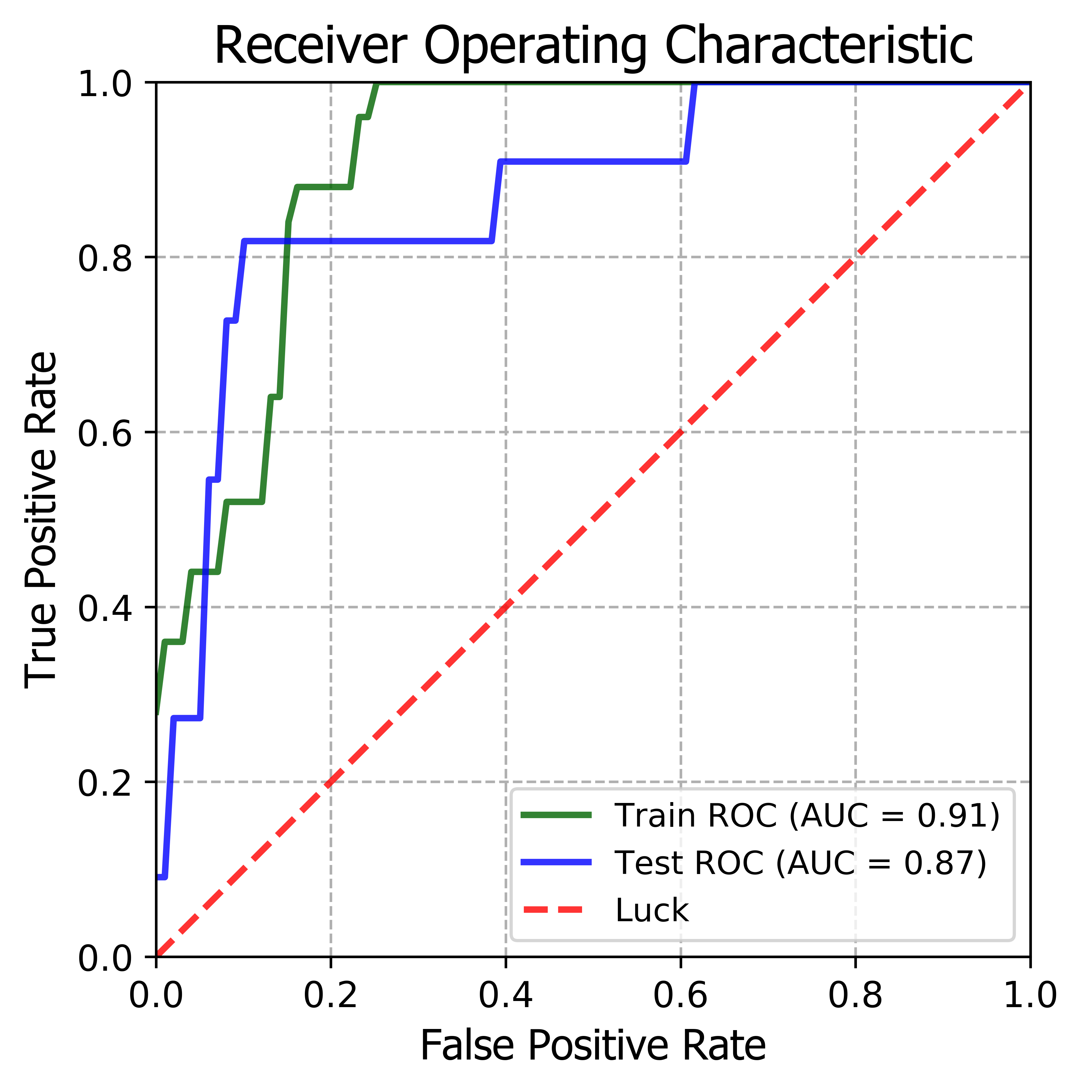
For liver function classification, 15 radiomics features were retained for constructing a prediction model, the overall accuracy and the mean AUC on cross-validation dataset (n=149) for prediction of Child-Pugh A is about 80.5% and 0.89±0.04 respectively. The model achieved AUCs of 0.91 and 0.87 on training and test datasets, respectively. . For prediction of liver cirrhosis from both normal liver and chronic hepatitis (HBV or/and HCV infection), 15 radiomics features were retained for constructing a prediction model, the overall accuracy and the mean AUC on cross-validation dataset (n=149) is about 83.2% and 0.90±0.06 respectively. By using the cutoffs, the AUCs were 0.93 and 0.93 in training and test datasets, respectively. Feature coefficients of trained model for predictions of liver function and liver cirrhosis were shown in Figure 2-3.The ROC curves were shown in Figure 4-5Conclusion
Radiomics analysis of gadoxetic acid-enhanced hepatobiliary phase images allows for accurate diagnosis of liver fibrosis. we used radiomics analysis of 2D-view whole liver at portal level on HBP MRI to investigate its value in prediction of Child-Pugh A and hepatic cirrhosis, our results showed a high diagnostic performance. Compared to reduced regular ROIs of CT or MR images in liver parenchyma for texture analysis(6,7), 2D-view of whole liver at portal level on HBP may be an optimal section to reflect the liver morphology information, enhancement of liver parenchyma and texture features, which may yields a better performance in diagnosing liver cirrhosis. Recently, Park HJ et al(8) reported a similar result that radiomics analysis of gadoxetic acid–enhanced hepatobiliary phase images allows for accurate diagnosis of liver fibrosis. They selected ROIs of the right hepatic lobe at the level of the right portal vein. However, the statistical power was limited due to the relatively small number of samples. Further research will be necessary to verify our preliminary findings in a larger cohort. Our promising results might encourage us to develop a convolutional neural network system to overall assess liver cirrhosis.Acknowledgements
References
1. Poynard T, Yuen MF, Ratziu V, Lai CL. Viral hepatitis C. Lancet 2003;362(9401):2095–2100.
2. Bravo AA, Sheth SG, Chopra S. Liver biopsy. N Engl J Med 2001;344(7):495–500.
3.Feier D, Balassy C, Bastati N, Stift J, Badea R, Ba-Ssalamah A. Liver fibrosis: histopathologic and biochemical influences on diagnostic efficacy of hepatobiliary contrast-enhanced MR imaging in staging. Radiology 2013;269(2):460–468.
4. Norén B, Forsgren MF, Dahlqvist Leinhard O, et al. Separation of advanced from mild hepatic fibrosis by quantification of the hepatobiliary uptake of Gd-EOB-DTPA.Eur Radiol 2013;23(1):174–181.
5. Venkatesh SK, Yin M, Takahashi N, Glockner JF, Talwalkar JA, Ehman RL. Noninvasivedetection of liver fibrosis: MR imaging features vs. MR elastography. Abdom Imaging 2015;40(4):766–775.
6. Lubner MG, Malecki K, Kloke J, Ganeshan B, Pickhardt PJ. Texture analysis of the liver at MDCT for assessing hepatic fibrosis. Abdominal radiology. 2017;42(8):2069-78.
7. Yokoo T, Wolfson T, Iwaisako K, Peterson MR, Mani H, Goodman Z, et al. Evaluation of Liver Fibrosis Using Texture Analysis on Combined-Contrast-Enhanced Magnetic Resonance Images at 3.0T. BioMed research international. 2015;2015:387653.
8.Park HJ, Lee SS, Park B, Yun J, Sung YS, Shim WH, et al. Radiomics Analysis of Gadoxetic Acid-enhanced MRI for Staging Liver Fibrosis. Radiology. 2019;292(1):269.
Figures