2474
A preliminary study on differential diagnosis of the benign and malignant liver lesions with amide proton transfer-weighted (APTw) imaging1The First Affiliated Hospital of Dalian Medical University, Dalian, China, 2Philips Healthcare, Beijing, China
Synopsis
Our work aimed to explore the value of amide proton transfer-weighted (APTw) imaging in diagnosing liver lesions. The result showed that novel imaging tool had a high value in differentiating benign liver lesions from malignant liver lesions (AUC: 0.801; sensitivity: 76.9%; specificity: 83.3%).
Introduction
Amide proton transfer-weighted (APTw) imaging is a novel MRI imaging tool based on endogenous substance in tissue, namely the amide protons in mobile cellular proteins and peptides[1]. Previous studies have shown that APTw imaging was applied to diagnosing central nervous system diseases and cervical Cancer[2,3]. To our knowledge, the application of APTw imaging in liver and related literature reports, especially the diagnosis of benign and malignant liver lesions, are rare.Materials and Methods
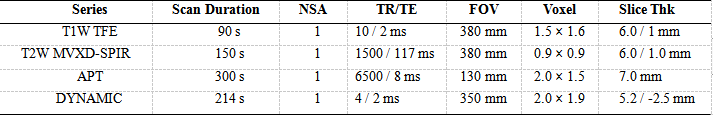
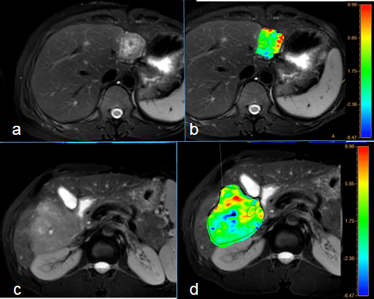
This study has been approved by the local IRB. 25 cases of focal liver lesions in our hospital were retrospectively analyzed,including 12 cases of benign lesions (4 males, 8 females, (52.36 ± 17.17) years old; hemangioma, n=11;FNH, n=1) and 13 cases of malignant lesions (12 males, 1 female, (59.92 ± 8.24) years old; hepatocellular carcinoma, n=9; Intrahepatic cholangiocellular carcinoma, n=4). The T1-wighted (T1w) turbo field echo (TFE), T2-weighted (T2w) with motion correction technique MultiVane XD (MVXD) and Spectral Presaturation with Inversion Recovery (SPIR) for fat suppression, diffusion weighted imaging (DWI), APTw and dynamic contrast enhanced (DCE) sequences were performed on a 3.0T MR scanner (Ingenia CX, Philips) (Table 1). Referring to the lesion information obtained on T2w images, circle ROIs were manually placed on the axial slice showing the largest liver lesions on APTw images by two radiologists independently. The average APTw values were calculated to minimize measurement bias. The consistency of APTw measurements of the lesions between the two radiologists was tested using intra-class correlation coefficients (ICC) in SPSS (IBM). APTw values were compared between the two groups using Independent-samples T test. ROC curve was drawn to analyze the parameter in the diagnostic efficiency of benign and malignant liver lesions.Results
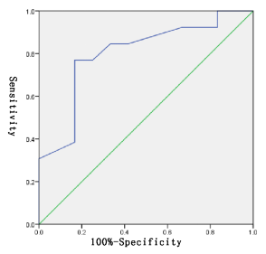
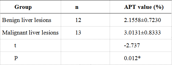
There was a good agreement between the two observers with ICC = 0.949. APT values of the benign and malignant lesions were (2.1558 ± 0.7230) % and (3.0131 ± 0.8333) %, respectively, with a significant difference (P = 0.012) (Table 2). Area under the curve (AUC) for APTw values to differentiate benign from malignant liver lesions was 0.801 and the feasible threshold was 2.485 with sensitivity of 76.9% and specificity of 83.3% (Figure 2).Discussion and Conclusion
Our study revealed that malignant liver lesions showed significantly higher APTw values than benign liver lesions, which was consistent in the previous studies for benign and malignant lesions arising from other anatomic sites[4,5].APTw imaging has a potential in the differential diagnosis of benign and malignant liver lesions. Malignant liver lesions showed significantly higher APTw values than benign liver lesions (AUC: 0.801; sensitivity: 76.9%; specificity: 83.3%). As a novel technology generating image contrast without the contrast medium, APTw imaging can provided a non-invasive approach for detecting benign and malignant liver lesions.Acknowledgements
No acknowledgement foundReferences
[1] Zhou J, Heo HY, Knutsson L et al. APT-weighted MRI: Techniques, current neuro applications, and challenging issues. Magn Reson Imaging, 2019, 50 (2): 347-364.
[2] Debnath A, Gupta RK, Singh A. Evaluating the Role of Amide Proton Transfer (APT)-Weighted Contrast, Optimized for Normalization and Region of Interest Selection, in Differentiation of Neoplastic and Infective Mass Lesions on 3T MRI. Mol Imaging Biol, 2019, undefined: undefined.
[3] He YL, Li Y, Lin CY, Qi YF et al. Three-dimensional turbo-spin-echo amide proton transfer-weighted mri for cervical cancer: A preliminary study. Magn Reson Imaging, 2019, 50 (4): 1318-1325.
[4] Yu L, Li C, Luo X, et al. Differentiation of Malignant and Benign Head and Neck Tumors with Amide Proton Transfer-Weighted MR Imaging. Mol Imaging Biol, 2019, 21 (2): 348-355.
[5] Ohno Y, Kishida Y, Seki S et al. Amide proton transfer-weighted imaging to differentiate malignant from benign pulmonary lesions: Comparison with diffusion-weighted imaging and FDG-PET/CT. J Magn Reson Imaging, 2018, 47 (4): 1013-1021.
Figures