2467
Measurement of pancreatic volume: a comparison between the single-layer ROI based and 3D volume Extraction methods.1Department of Radiology, the First Affiliated Hospital of Dalian Medical University, Dalian, China, 2Philips Healthcare, Beijing, China
Synopsis
It has been reported that pancreatic volume is closely related to pancreatic function. The 3D mDixon Quant MRI can achieve accurate water and fat images. Here, two measurement methods, including the single-layer region-of-interest (ROI) based and the 3D volume extraction methods, were compared for determination of pancreatic volume on the 3D mDixon Quant images. Results showed that pancreatic volumes measured by the two methods were with favorable consistency (ICC=0.971) and strong stability (P=0.169).
Introduction
Pancreas plays an important role in endocrine and exocrine functions of human body, and the function of pancreas can be associated with its volume[1-2]. Individuals with large pancreas are thought to have a big pool of B-cells that can better resist the development of diabetes. Therefore, quantitative detection of pancreatic volume can be of important clinical significance. As far as we know, application of the three-point Dixon sequence for determination of pancreas volume has been previously studied. The advanced mDixon Quant technique[3] adapts 6-echo fast 3D gradient echo acquisition and 7-peak reconstruction with T2* and novel eddy current compensation, therefore, achieves more accurate water and fat images. Here, using the 3D mDixon Quant images, two measurement methods, including the single-layer region-of-interest (ROI) based and the 3D volume extraction based methods, were compared for determination of pancreatic volume.Materials and methods
27 subjects (15 males, 12 females,age from 12 to 62 years old) were included, and underwent upper abdomen MRI scans (including 3D mDIXON Quant) on a 3.0 T MR scanner (Ingenia CX; Philips Healthcare, the Netherlands). This study has been approved by the local IRB. Scanning parameters for the 3D mDIXON Quant sequence were as:FOV = 375 × 300 × 168 mm³, TR/TE = 6.0/1.05 ms, flip angle = 3°, voxel size = 2.3 × 2.31 × 5.0 mm3, data matrix = 164 × 130 × 67, and scan duration = 17.9s. Pancreatic volumes were quantitatively measured by two observers with diagnostic experiences of 3 and 5 years, respectively, using double-blind methods(Figure 1-2). Measurement consistency between the two observers was evaluated with intra-class correlation coefficients (ICC). The Shapiro-Wilk test was used to check data normality of measurements by the two methods. The Mann Whitney test and ICC were respectively used to analyze the difference and consistence between measurements by the two methods.Results
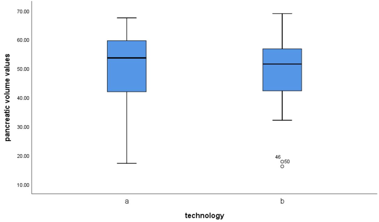
Measurements by the two observes were in good agreement (Figure 3). Since the pancreatic volumes measured by the single-layer ROI-based method were non-normally distributed, its average value was expressed as medians (25th, 75th percentiles) = 53.68 (37.49, 61.71) cm3; while for those measured by the 3D volume extraction method were in accordance with normal distribution, and its average volume was described as 48.37 ± 13.36cm3 (Figure 4). Pancreatic volumes measured by the single-layer ROI-based and 3D volume extraction methods on the 3D mDIXON Quant images show favorable consistency (ICC > 0.75) and strong stability (P > 0.05).Discussion and conclusion
In summary, both the single-layer ROI based and the 3D volume extraction methods can obtain accurate determination of the pancreatic volume. But the single-layer ROI method can obtain pancreatic fat fraction at mean time, which can be more practical than the 3D volume extraction method for clinical applications.Acknowledgements
No acknowledgements found.References
1. Sakata N, Egawa S, Rikiyama T, et al. Computed tomography reflected endocrine function of the pancreas. J Gastrointest Surg. 2011;15(3):525-532.
2. Williams AJ, Thrower SL, Sequeiros IM. Pancreatic volume is reduced in adult patients with recently diagnosed type 1 diabetes. J Clin Endocrinol Metab. 2012;97(11):2109-2113.
3. Macauley M, Percival K, Thelwall PE, et al. Altered volume, morphology and composition of the pancreas in type 2 diabetes. PLoS One. 2015;10(5):e0126825.
Figures
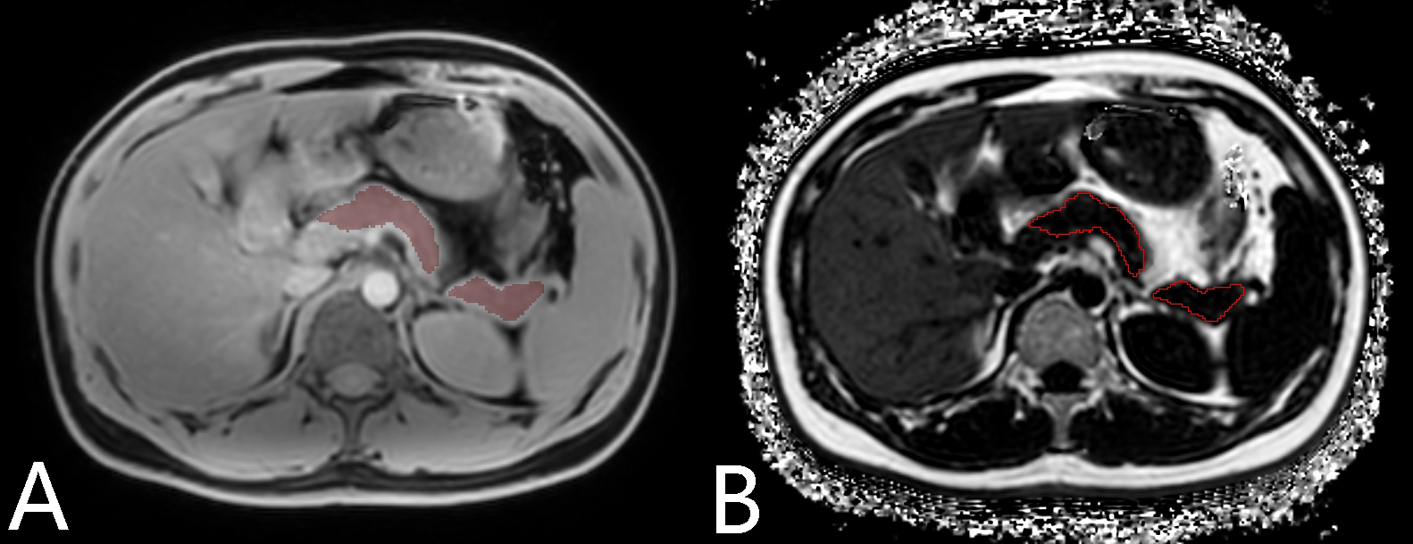
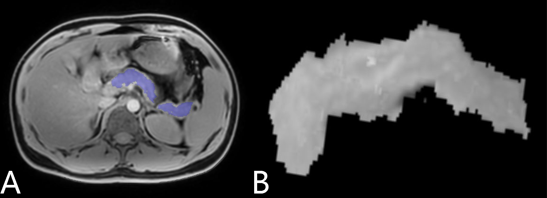
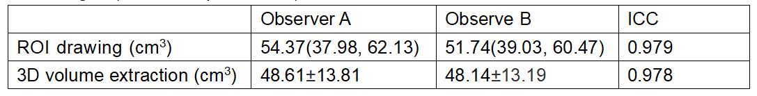
Fig.3 Pancreatic volume measurements by the two observers (A, B) using the singer-layer ROI drawing superposition volume and 3D volume extraction methods, and results for the intra-group consistency test. Normally distributed data were expressed as means ± standard deviations. Normally distributed data were expressed as medians and ranges (25th, 75th percentiles).