2455
Evaluate the efficiency of the ESWAN value for differentiating renal clear cell carcinoma with different pathological grades.1The First Affiliated Hospital of Dalian Medical University, dalian, China, 2GE Healthcare, Beijing, China
Synopsis
To investigate the potential of ESWAN in differentiating renal clear cell carcinoma with different pathological grades, to help preoperative non-invasive prediction of renal clear cell carcinoma pathological grading.
Synopsis
To investigate the potential of ESWAN in differentiating renal clear cell carcinoma with different pathological grades, to help preoperative non-invasive prediction of renal clear cell carcinoma pathological grading.INTRODUCTION
Renal cell carcinoma (RCC) constitutes approximately 3.8% of new cancers, in which clear cell renal cell carcinoma (ccRCC) is the most common type accounting for approximately 80% of RCC. The tumor stage and grade are the most important prognostic predictors of 5-year survival [1].Malignant tumor cells promote the formation of pathological neovascularization, but this neovascular structure is unstable, irregular in shape, and branch disorder, which is more likely to form arteriovenous malformations and is prone to rupture and hemorrhage. In addition, the metabolism of malignant tumors is stronger than that of normal cells, and it can ingest more nutrients such as sugar, protein, and oxygen, which together lead to the relative hypoxia of tumor tissues [2].The ESWAN sequence can quantitatively determine the degree of hypoxia in the tumor and detect microbleeds in the lesion according to the change of the magnetically sensitive paramagnetic substance,and potentially discriminate histological changes in kidneys non-invasively. [3] Previous studies have shown that ESWAN imaging has been used in the diagnosis of kidney disease. In this study, we aimed to investigate the potential of ESWAN in differentiating ccRCC with different pathological grades.METHODS
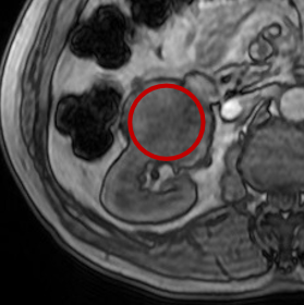
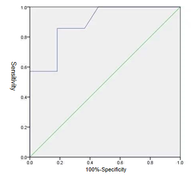
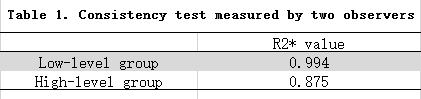
Patients with renal clear cell carcinoma who underwent abdominal MRI scan(GE Signa HDxt 1.5T MR) and ESWAN axial scan between January 2016 and October 2017 were retrospectively collected. These patients were confirmed by surgical pathological results, divided into low-level group (Fuhrman I ~ II) 11 cases, high-level (Fuhrman III ~ IV) 7 cases , a total of 18 cases.This study has been approved by the local IRB.Exclusion criteria: (1) Image artifacts are obvious, interference data measurement; (2)tumor diameter <1cm, the ROI error is large;(3)lesions are postoperative recurrence;(4)other pathological types of kidney tumors. All 18 volunteers were enrolled in the study eventually. After all data were transferred to the Data post-processing workstation,image analysis and data measurement were then performed using 4.6 software. Two radiologists with 2 and 5 years of MR diagnostic experience respectively performed the image measurements independently.The lesion R2* value measurement was performed using the Viewer function. At the largest level of the tumor, the ROI is placed according to the contour of the tumor,as shown in Figure 1.The average value of the ROI measured by the two observers was calculated for statistical analysis.The data were analyzed using SPSS version 23.0 with intraclass correlation coefficient (ICC) >0.75 indicating good consistencies. According to the data is in compliance with normality,Independent sample t test and Mann-Whitney U test were used to analyze the difference between the R2* values of ESWAN images obtained by the two observers. The receiver operating characteristic curve (ROC) was used to analyze the diagnostic performance,and the phase diagnostic efficacy AUC values and the corresponding sensitivity and specificity were calculated,as shown in Figure 2.
RESULTS
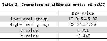
The consistency measured by two observers was good (ICC>0.75),as shown in Table 1. The R2* values of ESWAN images in high-grade RCC group were significantly higher than those in low-grade RCC group (shown in Table 2, P=0.028<0.05). And that obtained an area-under-curve (AUC) for ROC study of 0.89 with sensitivity of 85.7% and specificity of 81.8%.DISCUSSION AND CONCLUSIONS
In this study, The R2* values of ESWAN images was first applied to differentiate renal clear cell carcinoma with different pathological grades.Renal clear cell carcinoma has different proliferation rate and cell density, and there are corresponding differences in oxygen consumption and degree of hypoxia. When ccRCC provides blood angiogenesis and structural abnormalities, it can cause blood supply and hemodynamic changes. These factors can lead to changes in the concentration of paramagnetic substances such as ccRCC blood metabolites. The ESWAN sequence can quantitatively determine the degree of hypoxia in the tumor, detect microbleeds, etc., and has potential advantages in the diagnosis and differential diagnosis of ccRCC.Acknowledgements
Thanks for all my teachers.References
1.Jun S, Yongqiang T, Jingjing C. et al.Clear cell renal cell carcinoma: CT-based radiomics features for the prediction of Fuhrman Grade.European Journal of Radiology.S0720-048X(18)30355-3.
2.Pinato D J, Pai M, Reccia I, et al. Preliminary qualification of a novel, hypoxic-based radiologic signature for trans-arterial chemoembolization in hepatocellular carcinoma[J]. Bmc Cancer, 2018, 18(1):211.
3.Ji S, Zhang S, Mao Z, et al. Quantitative assessment of iron deposition in Parkinson’s disease using enhanced T2 star-weighted angiography[J]. Neurol India, 2016, 64(3): 428-35.
Figures