2453
Comparison of Diagnostic Performance of Magnetic Resonance Imaging and Computed Tomography for Evaluation of Complex Renal Cysts1Tongji Hospital, Tongji Medical College, Huazhong University of Science and Technology, Wuhan, China
Synopsis
To improve understanding of DWI features of complex renal cysts, and compare diagnostic performance of MRI and CT in discrimination of benign and malignant masses. Images of each lesion were analyzed, including size, thickness of wall, number of septum, enhancement of wall/septum, wall nodule, calcification, and cyst content. CT and MRI image characteristics were compared with pathology or follow-up results. The incidences of high signal intensity on DWI were significantly higher in malignant than in benign masses. MRI showed higher AUC than CT for differentiating benign from malignant masses. MRI could be useful in improving diagnosis of complex renal cysts.
Introduction/Purpose
Renal cystic masses are common and are being detected more and more frequently due to the increasing use of cross-sectional imaging. 21% of cystic masses show a complex pattern, making it difficult to identify benign or malignant and subsequent treatment1. At present, the Bosniak classification criteria based on CT has been widely used to evaluate renal cystic masses and guide clinical management2. However, due to the low contrast resolution of CT, the CT-based Bosniak classification criteria have certain limitations for detecting septa and solid components. In addition, there are some clinical factors and image findings that are helpful in identifying benign and malignant, such as body mass index (BMI) of patients and high signal intensity of solid components on diffusion weighted imaging (DWI), but are not included in the classification criteria. Therefore, the accuracy of Bosniak classification criteria only based on CT for the diagnosis of complex renal cysts remains controversial3. Studies have shown that the Bosniak criteria can also be useful on MRI4,5. The purpose of this study was to compare the diagnostic performances of CT and MRI in the evaluation of renal cystic mass according to pathology and follow-up results.Materials and Methods
This study retrospectively analyzed 85 renal cystic masses in 81 patients with CT or MRI, including 42 patients with DWI (b = 600, 1000 s/mm2). Clinical variables and histopathological results were recorded. Two radiologists in consensus analyzed images of each lesion for the size, thickness of wall, number of septum, enhancement of wall/septum, wall nodule, calcification, and cyst content. Clinical variables, CT and MRI image characteristics were compared with pathology or follow-up results to evaluate the diagnostic performance for renal cystic masses.Results
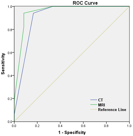
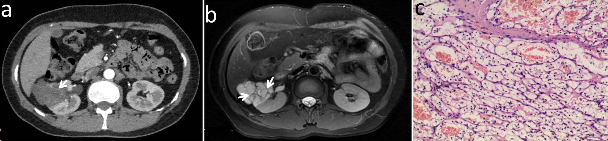
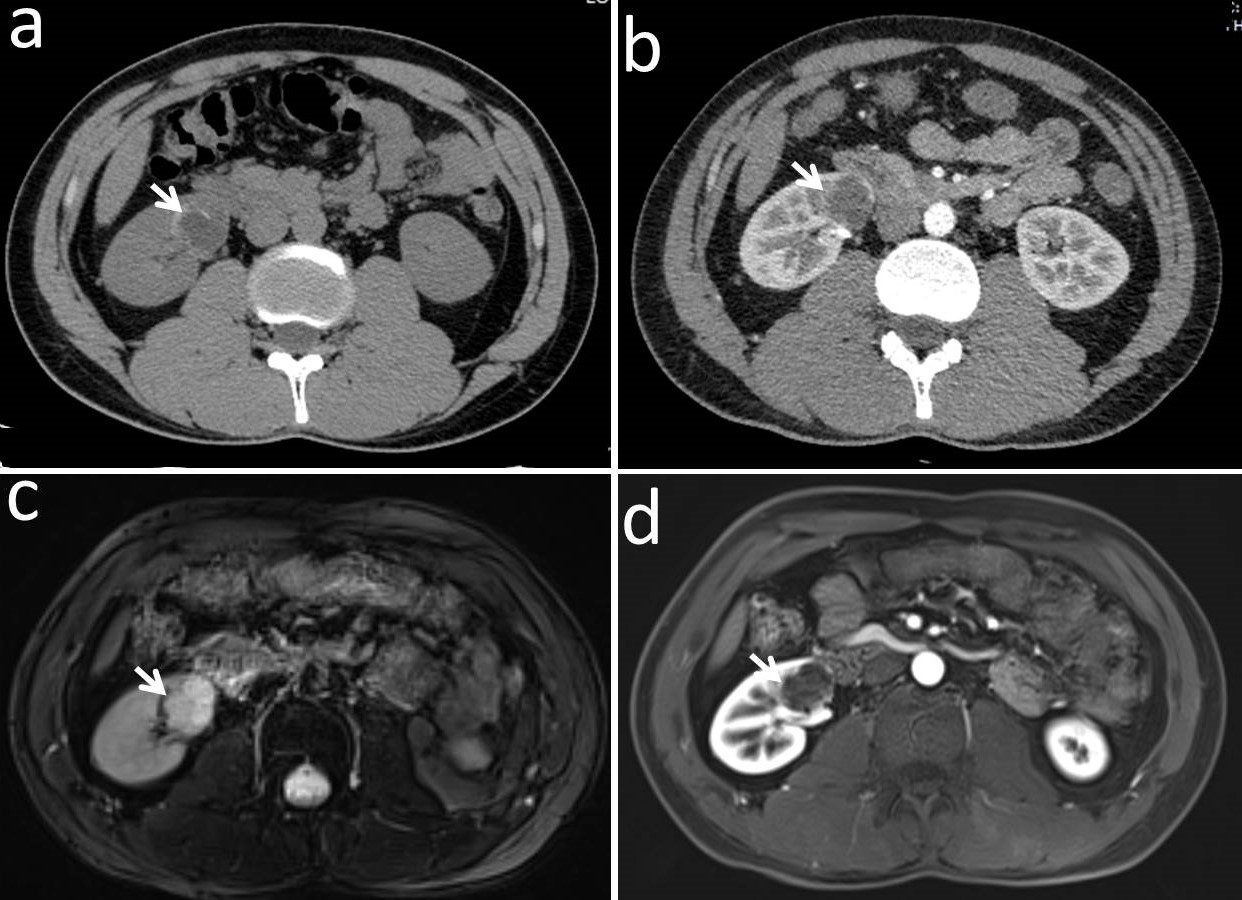
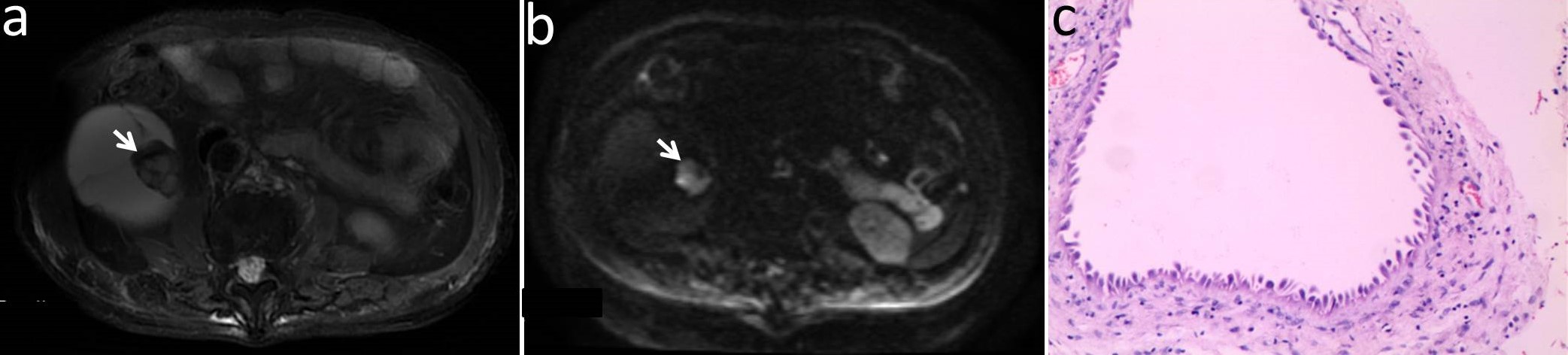
Of the 85 lesions in 81 patients, histological analysis reported that 31 were malignant, 31 were benign, and no change was found in 23 followed-up lesions (mean follow-up of 17 months). The incidences of cystic wall thickened, more septa, measurable enhancement of wall/septum, wall nodule on CT/MRI, and high signal intensity on DWI were significantly higher in malignant than in benign masses (CT: P = 0.007, P < 0.001, P = 0.001, P = 0.002, P < 0.001; MRI: P = 0.006, P < 0.001, P < 0.001, P = 0.020, P < 0.001, P < 0.001). MRI showed higher area under the receiver operating characteristic curve (0.951) than CT (0.912) for differentiating benign from malignant cystic masses (Fig. 1). The images of several representative cases are shown in Figure 2-4.Discussion
Because of the superiority in soft tissue resolution, MRI exhibits additional findings in some cases, such as irregular thickened walls or more septa4,6. In addition, DWI sequence of MRI can provide additional histological features inside the renal cystic masses6,7. Using contrast-enhanced MRI by subtraction imaging technique, the influence of calcification can be removed to determine whether it is really enhanced4,7. The diagnostic power of MRI for complex cysts was higher than that of CT in our study. This was similar to the previous research findings2-4,7. Because of the superiority in soft tissue resolution of MRI, the potential of DWI to detect malignant features, and enhanced MRI to avoid the effect of calcification, MRI may be more accurate and favorable than CT in evaluating renal cystic masses.Conclusions
MRI has high contrast resolution, functional imaging sequences such as DWI, lack of ionizing radiation, more sensitive depiction of septa and wall nodules, better characterization of enhancement, and more precise imaging of inner structure and content of the lesions. MRI could be useful in improving the assessment of complex renal cysts.Acknowledgements
No acknowledgement found.References
1. Li Y, Dai C, Bian T, et al. Development and prospective validation of a novel weighted quantitative scoring system aimed at predicting the pathological features of cystic renal masses. Eur Radiol 2018.
2. Sevcenco S, Spick C, Helbich TH, et al. Malignancy rates and diagnostic performance of the Bosniak classification for the diagnosis of cystic renal lesions in computed tomography - a systematic review and meta-analysis. Eur Radiol 2017;27:2239-47.
3. Defortescu G, Cornu JN, Bejar S, et al. Diagnostic performance of contrast-enhanced ultrasonography and magnetic resonance imaging for the assessment of complex renal cysts: A prospective study. Int J Urol 2017;24:184-9.
4. Israel GM, Hindman N, Bosniak MA. Evaluation of cystic renal masses: comparison of CT and MR imaging by using the Bosniak classification system. Radiology 2004;231:365-71.
5. Silverman SG, Pedrosa I, Ellis JH, et al. Bosniak Classification of Cystic Renal Masses, Version 2019: An Update Proposal and Needs Assessment. Radiology 2019;292:475-88.
6. Inci E, Hocaoglu E, Aydin S, Cimilli T. Diffusion-weighted magnetic resonance imaging in evaluation of primary solid and cystic renal masses using the Bosniak classification. Eur J Radiol 2012;81:815-20.
7. Pitra T, Pivovarcikova K, Tupy R, et al. Magnetic resonance imaging as an adjunct diagnostic tool in computed tomography defined Bosniak IIF-III renal cysts: a multicenter study. World J Urol 2018;36:905-11.
Figures