2450
Initial experience in abbreviated T2-weighted Prostate MRI using a Deep Learning reconstruction.1Massachusetts General Hospital, Boston, MA, United States, 2Applications and Workflow, GE Healthcare, Boston, MA, United States, 3Applications and Workflow, GE Healthcare, Calgary, AB, Canada, 4Applications and Workflow, GE Healthcare, Houston, TX, United States
Synopsis
Increasing the speed of multiparametric prostate MRI (mpMRI) is highly desirable. However, usual tradeoffs between signal-to-noise (SNR), scan time and lesion conspicuity must be considered. One recently proposed approach consists of using a bi-parametric protocol, whereby only the T2 and diffusion-weighted images are collected, thus highlighting the particular significance of achieving a robust, high-quality T2-weighted acquisition. As such, this work focuses on evaluating a Deep Learning reconstruction technique which shows promises to cut acquisition time of prostate T2-weighted imaging in half and would therefore benefit both bi-parametric as well as mpMRI.
INTRODUCTION
Multiparametric Prostate MRI (mpMRI) requires tradeoffs between Signal-to-Noise Ratio (SNR), spatial resolution and scan time. A complete mpMRI examination at 3T can still take up to 25 min of gradient time even after optimizing protocols using commercially-available acceleration techniques. One recently proposed approach consists of using a bi-parametric protocol, whereby only the T2 and diffusion-weighted images are collected, thus highlighting the particular significance of achieving a robust, high-quality T2-weighted acquisition [1]. Recently, the application of Deep Learning to MR image reconstruction (DL Recon) has ushered in new acceleration strategies. Indeed, T2W prostate imaging is typically performed with multiple averages to achieve the highest spatial resolution at the expense of increased vulnerability to motion and additional scan time. In this study, we explore the possibility of leveraging DL Recon to speed up T2W acquisition beyond the capabilities of parallel imaging alone.METHODS
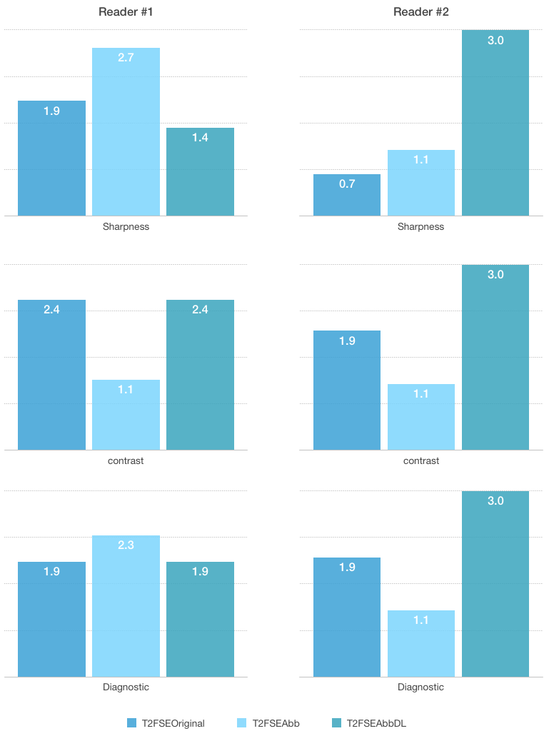
14 patients were scanned using our institution's standard mpMRI clinical protocol which includes an axial T2W Fast Spin Echo - referred to as T2FSEOriginal - (NEX=2.5; ETL=18; Matrix size: 320x228; FOV=20x20cm2; Slice thickness=4mm; TR/TE=3000/119ms; Total Scan Time=5min6s) on a GE Signa Premier 3T MRI system (GE Healthcare, Waukesha, WI, USA). The protocol was modified to accommodate an abbreviated T2W FSE - referred to as T2FSEAbb - collected with the exact same parameters as T2FSEOriginal, except that the number of NEX was set to 1 (Total Scan Time=2min6s). The T2FSEAbb raw data signals were then reconstructed in two ways, first using the product 2D cartesian reconstruction and second using a DL Recon algorithm based on a Convolutional Neural Network (CNN) trained to optimize SNR. For this third series referred to as T2FSEAbbDL, images were reconstructed with a tunable noise reduction factor set to 75%. The resulting 3 series were independently reviewed by two board-certified abdominal radiologists who were asked to rank them according to the following image quality criteria: sharpness, absence of artifacts, perceived contrast, most diagnostic. The readers were also asked to annotate images where pathology could have been missed due to inadvertent conspicuity differences across the 3 series.RESULTS
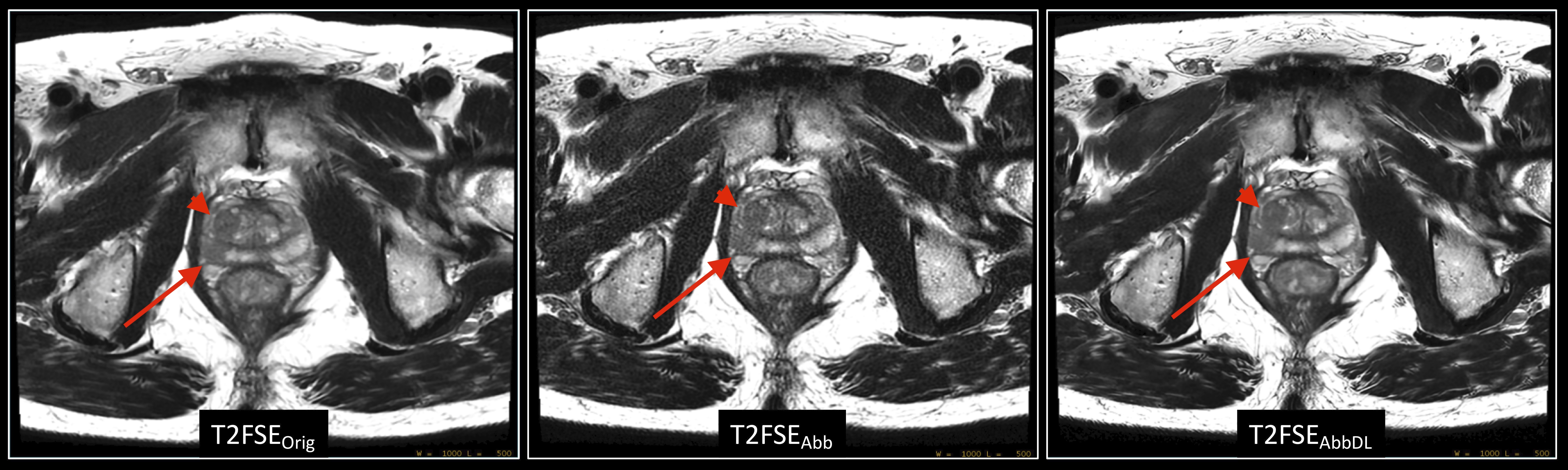
The results are summarized in Figure 1. The T2FSEAbbDL series was assessed to have better sharpness, perceived contrast and diagnostic value by Reader #2, whereas the rating from Reader #1 was more heterogeneous. Importantly, significant pathology was reported in 3 out of the 14 cases and was consistently detected across all 3 series. In one case, the Transition Zone (TZ) lesion was found by one reader to exhibit more clinically concerning features in the T2FSEAbbDL series compared to the T2FSEAbb series. Biopsy results were not yet available to correlate with this finding. In another case, pulsation-related motion artifacts near the rectum impacted the single NEX series more than the T2FSEOriginal series. Conversely, severe motion artifacts found in a 2.5NEX series were otherwise not observed in the single NEX scan. No significant artifact (blurring, ringing, lack of SNR, etc) was found in the T2FSEAbbDL compared to the 2 series generated with conventional reconstruction. Figure 2 shows representative T2W images with the original and abbreviated series along with the series reconstructed with DL Recon in a patient with suspected focus of prostatic malignancy.DISCUSSION/CONCLUSION
In this multi-reader study, the proposed abbreviated T2FSEAbbDL scan offers a clinically viable method to reduce scan time by up to 50% while retaining sufficient SNR thanks to DL Recon. A current limitation of this on-going study is the relatively small cohort (N=14). However, these encouraging results warrant a continuation of the study and correlation with biopsy results, multiple readers and the utilization of image analysis metrics to address subjective bias [2]. In conclusion, DL Recon offers a new strategy to reduce scan time in mpMRI of the prostate by alleviating the need to collect multiple averages for T2W acquisitions.Acknowledgements
No acknowledgement found.References
[1] Sherrer, R.L. et al. Comparison of biparametric MRI to full multiparametric MRI for detection of clinically significant prostate cancer. Prostate Cancer Prostatic Diseases 22, 331–336 (2019) [2] Burgess, A.E. Visual Perception Studies and Observer Models in Medical Imaging. Seminars in Nuclear Medicine 41, 419–436 (2011)
[2] Burgess, A.E. Visual Perception Studies and Observer Models in Medical Imaging. Seminars in Nuclear Medicine 41, 419–436 (2011)
Figures