2448
Highly accelerated prostate water/fat 3D MRI with compressed sensing and SENSE, compared to the standard of care1The First Affiliated Hospital of Dalian Medical University, Da lian, China, 2Philips Healthcare,China, Bei jing, China
Synopsis
Compressed sensing(CS)technology is currently used less in prostate diseases. This study aims to explore the feasibility of CS and SENSE technology with different acceleration factors for prostate 3D magnetic resonance imaging(MRI). The preliminary exploration concluded that the SNR and CNR with CS 5 in prostate 3D mDIXON imaging are similar to the SENSE 2, and the imaging time is reduced by 28%, which effectively improves the MR scanning efficiency.
Purpose
To explore the effects of CS and SENSE techniques on prostate 3D mDIXON imaging with different acceleration factors.Introduction
Prostate disease is a common disease in elderly men, and the incidence rate has increased in the past years [1]. MRI has become the preferred imaging method for the diagnosis of prostate diseases due to its excellent soft tissue resolution, multi-parameter, multi-sequence and multi-directional imaging features [2]. Traditional MRI scans take a long time and some patients cannot tolerate them. Recently, the application of CS technology has improved the signal acquisition efficiency of 3D sequences and greatly shortened the imaging time [3]. However, there are few clinical studies using this technology. Therefore, this study explores the feasibility of different acceleration factors of CS and SENSE technology for prostate 3D MRI.Materials and Methods
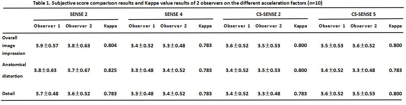
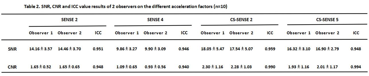
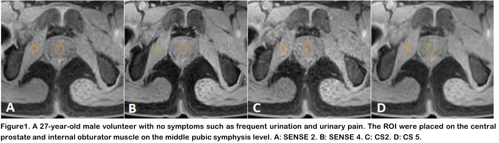
Ten healthy volunteers (mean 27.3 years) were prospectively involved for 3.0T MRI (Ingenia CX, Philips) with 3D GRE sequences (6-point Dixon water/fat) on prostate. A clinical routine protocol was scanned with SENSE factor 2, followed by a SENSE 4 to stretch the acceleration capability. CS with acceleration factors 2 and 5 were then prescribed to approximately match the scan times with SENSE 2 and 4. Other parameters were kept same: TR/TE 3.7/1.3 ms, resolution 1.5×1.7×3.00 mm3. Scan times were 17.7s, 11.2s,15.5s and 12.7s for SENSE 2, SENSE 4, CS 2, and CS 5. All image analysis was performed independently by two radiologists on ISP workstation (Philips), blind to the imaging sequence. ROIs with fixed area were placed as in Figure 1 for SNR and CNR calculations. These two radiologists also scored on the overall image impression, anatomical distortion and detail display according to Likert 5, which were analyzed for consistency using Kappa coefficient. The SNR and CNR were analyzed for consistency using intraclass correlation coefficient (ICC). The SNR and CNR of images among four imaging protocols were tested using Friedman-test. This study has been approved by the local IRB.Results
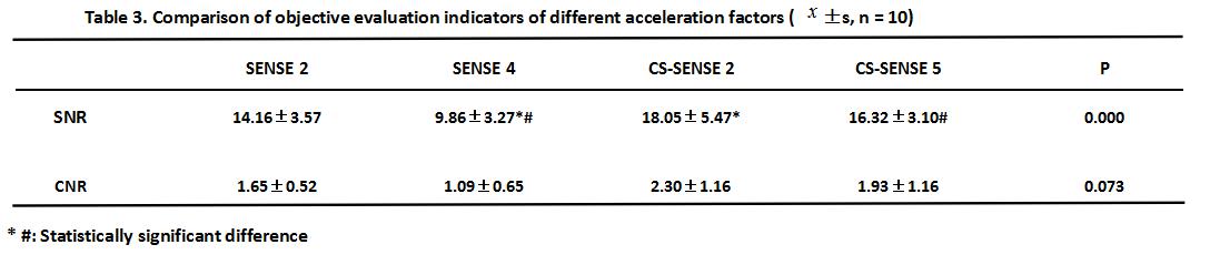
The SNR, CNR, and subjective scoring agreed well between the two radiologists (Kappa>0.75, ICC>0.75, Tables 1&2), and the data from the senior observer was chosen for the final statistical analysis. The average scores among these different acceleration factors were 3.8, 3.3, 3.5 and 3.5, without statistical difference (P=0.375). There was no statistical difference in SNR between CS 2 and CS 5, nor between CS5 and SENSE2. The SNRs of CS 2 and 5 were both higher than that of SENSE 4 (Table 3). There was no significant difference in CNR among the four protocols.Discussion and Conclusion
To our best knowledge, this is a preliminary study of applying the different acceleration factors of CS and SENSE technology for prostate 3D MRI. It is concluded that CS 5 yielded similar SNR and CNR in prostate water/fat imaging to those of SENSE 2, the clinical routine, while the imaging time was cut by 28%. CS with acceleration factor 5 is demonstrated a potential replacement for the clinical routine protocol for prostate water/fat imaging, given the retained image quality and the reduced imaging time, and these benefits are expected to increase with further improved resolution.Acknowledgements
No acknowledgement found.References
[1]GUO J D. The value of multi-parameters magnetic resonance imaging in the diagnosis of early prostate cancer and prostatitis in the peripheral zone. Modern diagnosis and treatment. 2019; 30 (9).
[2]ZHOU L, ZHOU X, ZHANG H W, et al. Comparative study of 3.0T magnetic resonance prostate T2WI PROPELLER and T2WI FSE. Journal of medical imaging. 2019; 29 (5).
[3]WANG Y K, JIN X, YUAN H S. Compressed sensing three-dimensional fast spin echo sequence for diagnosis of knee joint cartilage injury. Magnetic resonance imaging. 2019; 10 (5).
Figures