2445
Assessment of aggressiveness of prostate cancer: correlation of MRI ADC map textures with Gleason grade after radical prostatectomy1Tongji Hospital,Tongji Medical College, Huazhong University of Science & Technology, Wuhan, China
Synopsis
Prostate cancer is the 2nd leading diagnosed cancer in men worldwide. Radiomics extracts large amounts of quantitative image features from radiologic images and selects stable and clinically relevant radiomics biomarkers for disease assessment. 90 patients underwent mpMRI before radical prostatectomy, with subsequent pathologic evaluation. The texture features were extracted by the python-based pyradiomics package. The AUC of the model was 0.841, with sensitivity 69.6% and specificity 91.2%, which was significantly higher than mean ADC value or single texture feature. MRI ADC map texture evaluation may facilitate noninvasive assessment of aggressiveness of prostate cancer.
PURPOSE: To retrospectively investigate whether the MRI ADC map textures of prostate cancer correlates with the Gleason grade at pathologic evaluation after radical prostatectomy.
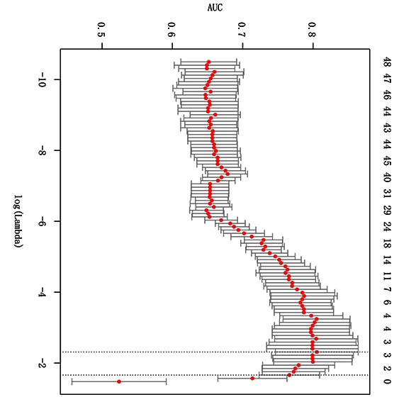
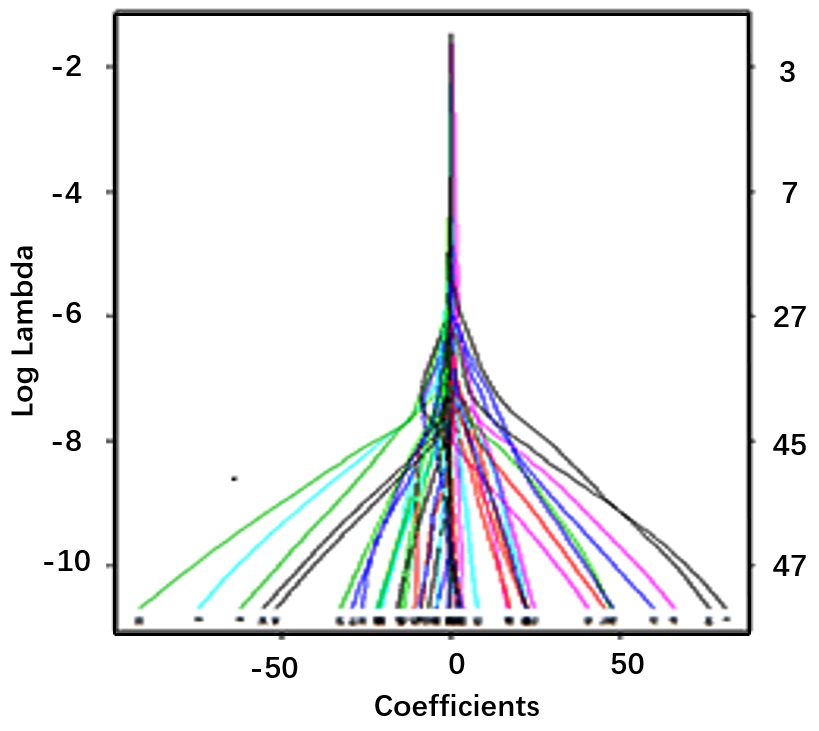
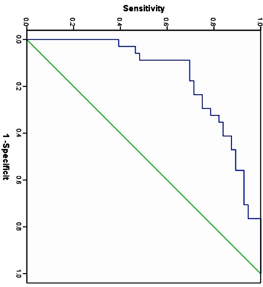
MATERIALS AND METHODS: The institutional review board approved and issued a waiver of informed consent for this study of 90 patients (age, 66.6±7.4 years) who underwent multiparametric magnetic resonance imaging (mpMRI) including T1WI,T2WI, DWI, and PWI, before radical prostatectomy, with subsequent pathologic evaluation, between May 2015 and June 2017.Inclusion criteria were that the patients had: no previous treatment including hormonal, irradiation, cryotherapy, or surgery; poor quality of the MRI images due to susceptibility artifacts, movement artifacts, or the presence of hip implants; and an interval of more than four weeks between mpMRI and biopsy. The ITK-SNAP software was used to manually drawn ROIs to encompass the whole tumor on the ADC maps. The texture features were extracted by the python-based pyradiomics package, including histogram features, GLCM, GLCM, GLDM, GLSZM, GLSZM and shape features. The repeatability of the texture features was evaluated with the ICC. Mann-Whitney U test and independent sample t-test were used to exclude features that had no significant difference between high grade (GS≥4+3) and low grade (GS≤3+4) prostate cancer. 5 fold cross-validation and Lasso regression model were used to obtain texture feature combination of the highest performance and develop a classification model for discriminating high grade (GS≥4+3) and low grade (GS≤3+4) prostate cancer. The diagnostic efficiency of the model was evaluated with ROC curve.
RESULTS:The area under the ROC curve (AUC) of the model was 0.841, with sensitivity 69.6% and specificity 91.2%, which was significantly higher than mean ADC value or single texture feature (P<0.05).
CONCLUSION: MRI ADC map texture evaluation may facilitate noninvasive assessment of aggressiveness of prostate cancer.
[Key words] Deep learning; Radiomics; Magnetic resonance imaging; Texture; Neoplasm grading
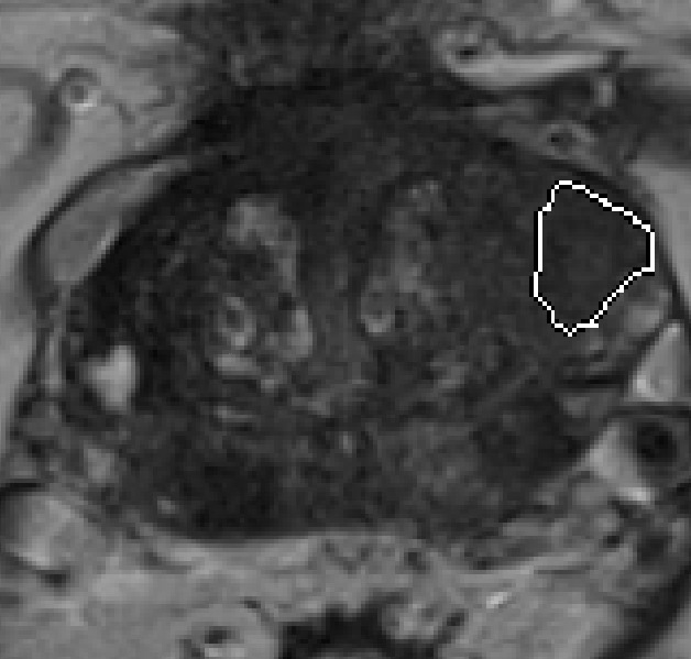
- T2WI (Fig.1 )
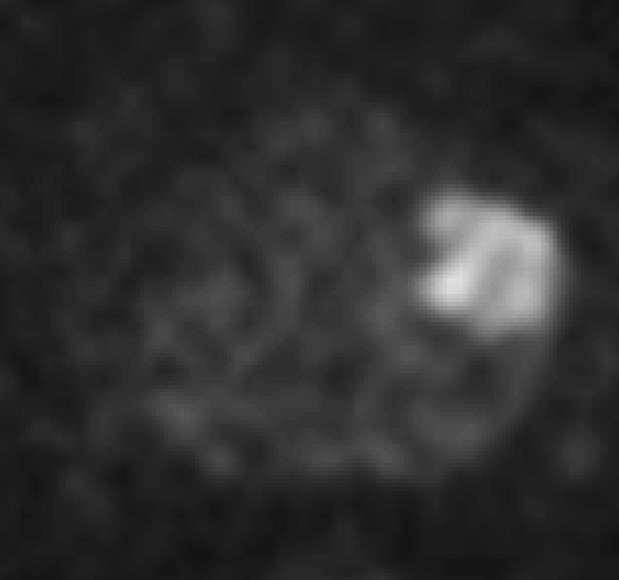
- DWI (Fig.2 )
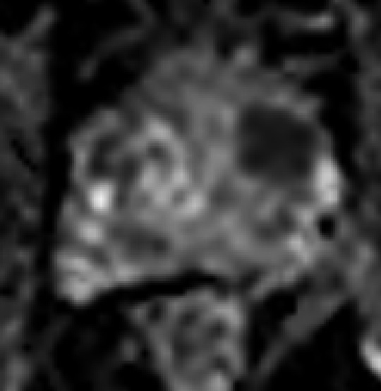
- ADC (Fig.3 )
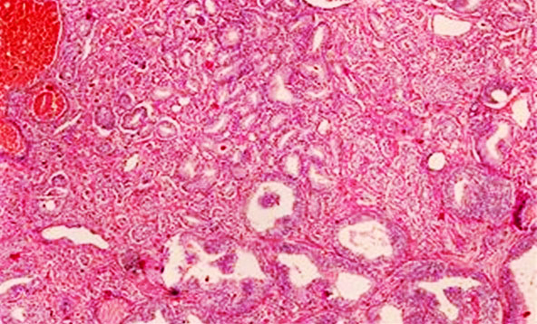
- Pathology Gleason 4+4,PSA 8.151 (Fig.4)
- 5 folds cross validation test (Fig.5)
- LASSO coefficient profiles (Fig.6)
- ROC curve (Fig.7)
Acknowledgements
This work was supported by the National Natural Science Foundation of China (81171307, 81671656)References
1. Bray F, Ferlay J, Soerjomataram I, et al. Global cancer statistics 2018: GLOBOCAN estimates of incidence and mortality worldwide for 36 cancers in 185 countries[J]. CA Cancer J Clin. 2018 Nov;68(6):394-424. PMID:30207593.doi: 10.3322/caac.21492
2. Thrall JH, Li X, Li Q,et al.Artificial Intelligence and Machine Learning in Radiology: Opportunities, Challenges, Pitfalls, and Criteria for Success[J].J Am Coll Radiol. 2018 Mar;15(3 Pt B):504-508. PMID: 29402533.doi: 10.1016/j.jacr.2017.12.026.
3. Smith CP, Czarniecki M, et al.Radiomics and radiogenomics of prostate cancer[J]. Abdom Radiol (NY). 2018 Jun 20. PMID:29926137.doi: 10.1007/s00261-018-1660-7.
4. Stoyanova R, Takhar M, Tschudi Y,et al. Prostate cancer radiomics and the promise of radiogenomics[J].Transl Cancer Res. 2016 Aug;5(4):432-447.PMID: 29188191
5. Abdollahi H, Mahdavi SR, Mofid B, et al. Rectal wall MRI radiomics in prostate cancer patients: prediction of and correlation with early rectal toxicity[J]. Int J Radiat Biol. 2018
Figures