2443
Differential diagnosis of Prostatic Hyperplasia with inflammation and Low-grade Transitional Zone Carcinoma: Advanced DWI VS Conventional DWI1Baoji Central Hospital, Baoji, China, 2GE Healthcare China, Beijing, China
Synopsis
The purpose of this study is to evaluate the efficacy of advanced diffusion weighted imaging (DWI) in the differential diagnosis of benign prostatic hyperplasia(BPH) with inflammation and Low-grade prostate transitional zone (TZ) cancer. By comparing the D, DDC and MK from advanced DWI with ADC value, it is found that the diagnostic efficiency of advanced DWI is better than the conventional DWI, and the combined diagnosis of (D + DDC + MK) has the highest efficiency.
Introduction
The differential diagnosis of benign prostatic hyperplasia (BPH) and prostate transitional zone (TZ) cancer is a daily work that clinicians have to perform. However, it is often difficult to differentiate TZ cancer from BPH, especially those BPH with inflammation and low-grade TZ cancer.. Previous studies reported that the BPH with inflammation demonstrated similar MR radiological characteristics to that of prostate TZ cancer, and the apparent diffusion coefficient ( ADC ) from the two lesions also overlaps1. In recent years, many advanced DWI models have been developed, including bi-exponential intravoxel incoherent motion (IVIM) DWI model, stretched exponential DWI model (SEM) and non-Gaussian diffusion kurtosis model (DKI). Many of these advanced DWI parameters are proven to be able to provide complementary information. The purpose of this study is to compare the diagnostic efficacy of conventional DWI and advanced DWI in the differential diagnosis of BPH with inflammation and low-grade TZ cancer, and to explore whether advanced DWI outperform conventional DWI in the differential diagnosis of these two diseases.Material and Methods
With pathologically confirmation from prostatectomy, from October 2017 to June 2019, 28 cases of BPH with inflammation (age 64.1 ± 4.2 years) and 29 cases of low-grade TZ carcinoma (age 71.2 ± 7.3 years) joined this study with written informed consents after the approval of the local Ethics Committee. In the prostate cancer group, 7 cases were Gleason Score (GS) = 3 + 2, 9 cases were GS = 3 + 3, and 13 cases were GS = 3 + 4. All patients were scanned on a 3.0T MR scanner (MR750w, GE Healthcare, Milwaukee, WI, USA) with a 16-channel body phased array coil. Imaging sequences included T1WI, T2WI, conventional DWI, Multi b-value DWI (for bi-exponential and stretched-exponential model fitting) and diffusion kurtosis imaging (DKI). Detailed scanning parameters were summarized in Table 1. The image post-processing and parameter measurement were carried out on a vendor provided Advanced Workstation (AW4.6 GE Healthcare). With reference of pathological results, multiple regions of interest (ROI) were delineated at the maximum level of the lesions. The ROI area was 30-150mm2, and the average value was calculated. The measured parameters include ADC value from traditional DWI, molecular diffusion coefficient (D) from IVIM model, distributed diffusion coefficient (DDC) from SEM and mean kurtosis (MK) from DKI. The difference of the parameters between the two groups was compared by independent sample t test, and AUC was calculated by ROC curve. The AUC of D, DDC , MK ,D + DDC + MK and ADC was compared by Z test.Results
The ADC, D and DDC values of BPH with inflammation were significantly higher than those of low-grade TZ carcinoma (P < 0.05), while the MK value was significantly lower(P < 0.05) (Figure 1, table 2). D, DDC, MK and combined parameter (D + DDC + MK) all have higher diagnostic efficacy compared with ADC (0.902, 0.904, 0.994, 0.966 vs 0.725, P < 0.05), among which the AUC of combined parameter (D + DDC + MK) is the highest. Although the diagnostic efficacy is not statistically different from D value, DDC value and MK value, the specificity of diagnosis is significantly improved. (Figure 2, table 3).Discussion and Conclusion
There are few studies on MR differential diagnosis of BPH with inflammation and Low-grade TZ carcinoma. When BPH is accompanied by inflammation, the inflammatory cells will aggravate the destruction of glands, lead to hyperplasia and fibrosis of glands, restrict the movement of local water molecules, and make them overlap with ADC value of TZ carcinoma2. Previous studies have shown that D value, DDC value and MK value can more easily detect the difference of decreased water molecule diffusion between BPH and TZ cancer than ADC value3,4. The results of this study is consistent with them in terms of D, DDC and MK from advanced DWI outperform ADC from conventional DWI in the differential diagnosis of Hyperplastic inflammation and Low-grade TZ cancer. Furthermore, combining D, DDC and MK provide the best diagnostic sensitivity and specificity in our study.Acknowledgements
References
1.Lee S-M, Wolfe K, Acher P, Liyanage SH. Multiparametric MRI appearances of primary granulomatous prostatitis. Br J Radiol 2019; 92(1098): 20180075.
2.Xiaohang L, Bingni Z, Liangping Z,et al. Differentiation of prostate cancer and stromal hyperplasia in the transition zone with histogram analysis of the apparent diffusion coefficient[J]. Acta Radiologica,2017,58(12): 1528-1534.
3.Liu X,Zhou L,Peng W,et al. Comparison of stretched-Exponential and monoexponential model diffusion-weighted imaging in prostate cancer and normal tissues. J Magn Reson Imaging,2015,42(4):1078-1085.
4.Mazzoni L N,Lucarini S,Chiti S,et al. Diffusion‐weighted signal models in healthy and cancerous peripheral prostate tissues: Comparison of outcomes obtained at different b‐values[J]. Journal of Magnetic Resonance Imaging,2014,39(3):512-518.
Figures
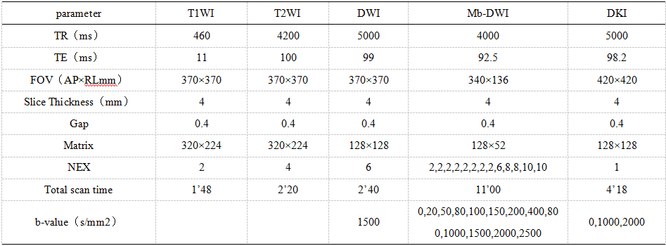
Table 1:MRI sequence scanning parameters
B value of IVIM-DWI sequence in mb-DWI is 0-1000 s/mm2, and b value of DWI sequence in tensile index model is 0-2500 s/mm2.
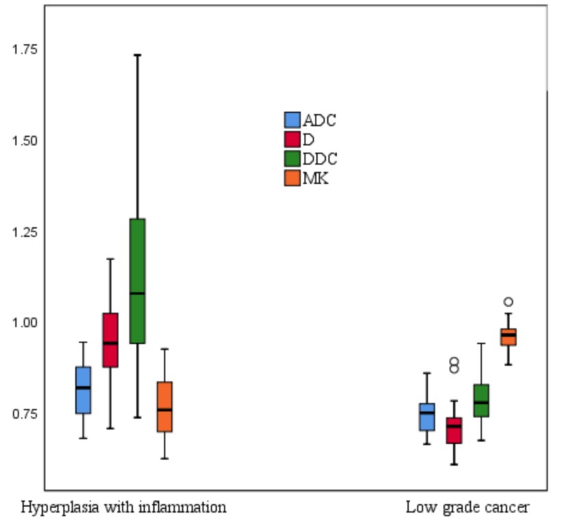
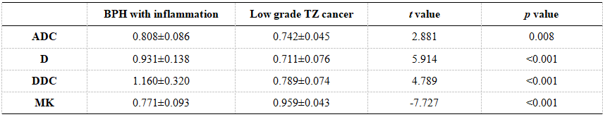
Table 2:Comparison of conventional ADC value, D value, DDC value and MK value between BPH with inflammation and Low-grade TZ cancer
ADC, D, and DDC, are in units of 10−3mm2/s.
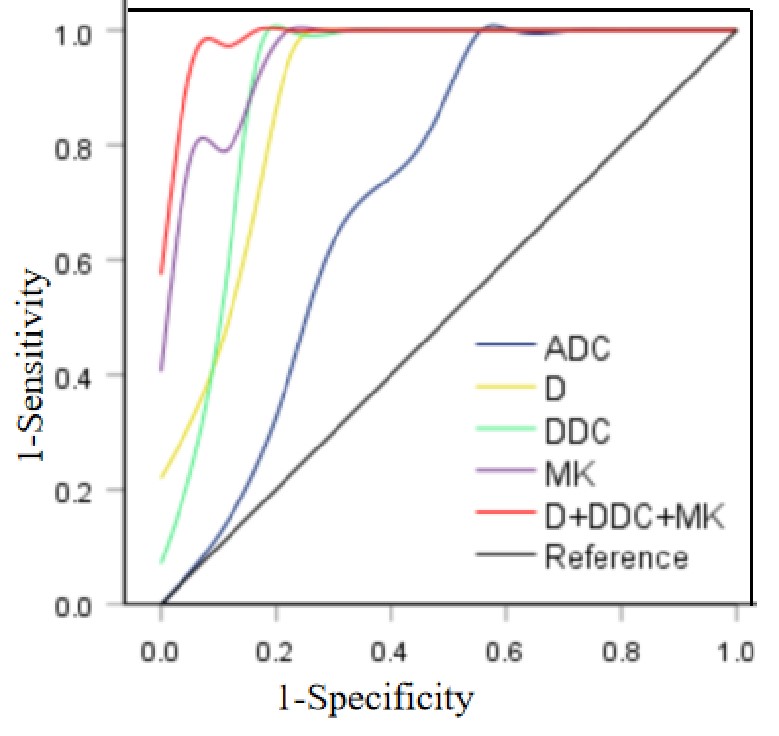
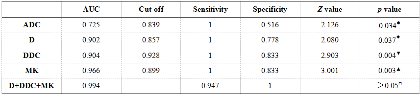
Table 3:Comparison of diagnostic efficacy of conventional DWI and advanced DWI in the differential diagnosis of BPH with inflammation and Low grade TZ cancer
The cut-off values of ADC, D, and DDC, are in units of 10−3 mm2/s; ●:ADC vs D; ◆:ADC vs DDC; ▼:ADC vs MK; ▲:ADC vs D+DDC+MK; □:D+DDC+MK vs D、DDC、MK.