2437
Amide Proton Transfer Imaging of Rectal Cancer: Which Duration and Power Level of Saturation Pulse is better?1Union Hospital, Tongji Medical College, Huazhong University of Science and Technology, Wuhan, China
Synopsis
We speculate that APT value may be a useful biomarker for assessing rectal pathological characteristics, which could have a potential impact on the clinical therapeutic strategies for patients. A baseline of APT value needs to be established for rectal cancer, which remains challenging due to the susceptibility effects. So we need to optimize the sequence by adjusting duration and power level of saturation pulse
INTRODUCTION
Rectal cancer is usually very proliferative and possesses high considerable potential in long-distance metastases [1], which likely lead to an abundant pool of mobile cellular proteins and peptides. We speculate that APT[2] value may be a useful biomarker for assessing rectal pathological characteristics, which could have a potential impact on the clinical therapeutic strategies for patients. A baseline of APT value needs to be established for rectal cancer, which remains challenging due to the susceptibility effects. So we need to optimize the sequence by adjusting duration and power level of saturation pulse.METHODS AND MATERIALS
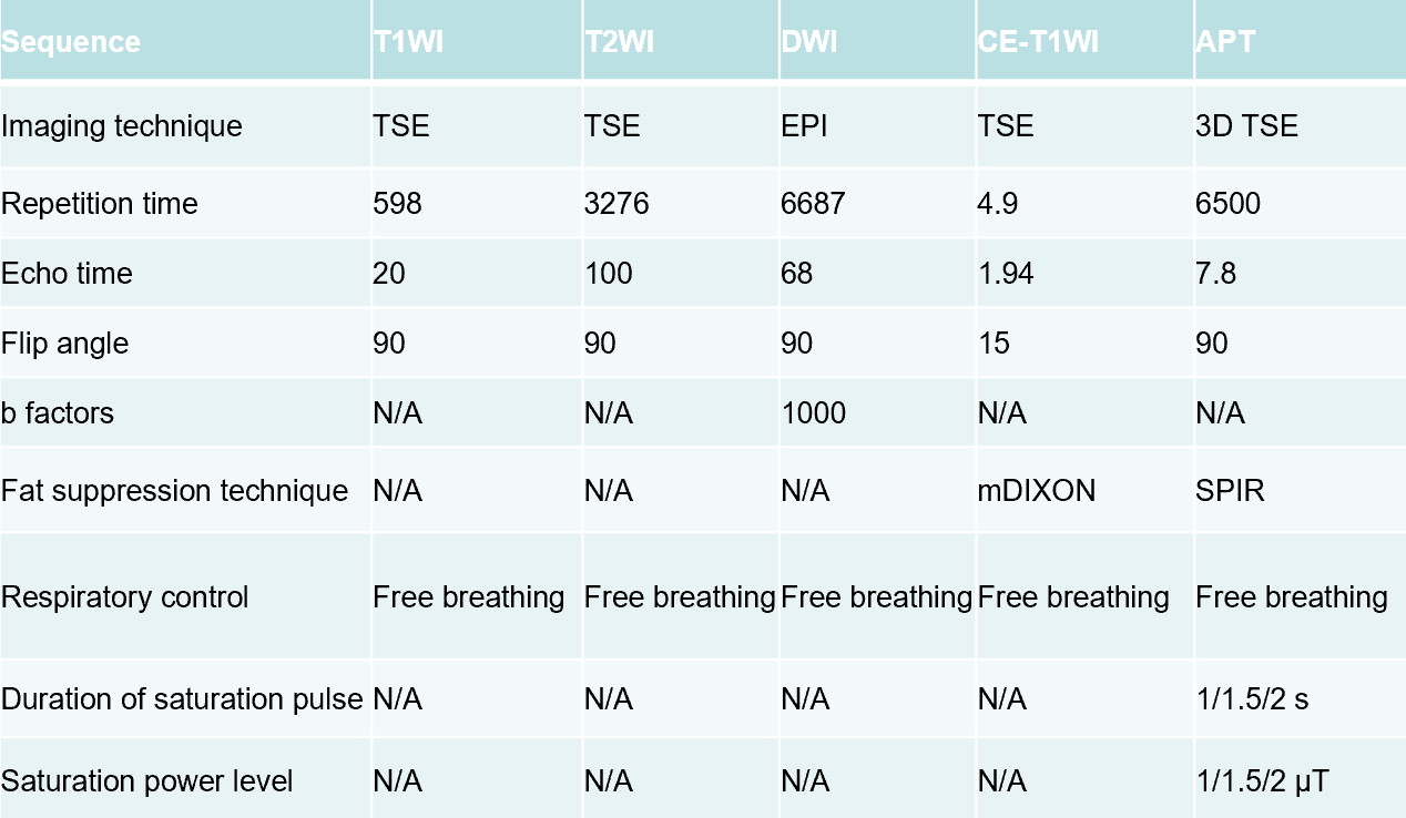
Five patient with locally advanced rectal cancer (LARC) were involved into this institutional review board-approved study. All participants were scanned at 3.0T (Ingenia CX, Philips Healthcare, Best, the Netherlands) using standard rectal protocols (T1WI, T2WI, DWI, contrast-enhanced) and APTw-imaging (including 7 groups of duration and power level of saturation pulse, acquired with 3D turbo-spin-echo sequence), imaging parameters listed in Table 1 and table 2. APT values [3] were measured by two radiologists independently drawing ROIs on a commercially available post-processing workstation (IntelliSpace Portal, Philips Healthcare, Best, the Netherlands). Polygonal ROIs were placed in the solid component of a tumor on the T2WI image, avoiding cystic, large necrotic, or hemorrhagic components, and copied onto the APTw image by each radiologist. The size of ROIs were draw ≥260 mm2 (≥80 pixels) and as large as possible, with exemplary ROIs shown in Figure 1. The imaging assessment was based on a 5-point scale by 2 radiologists independently, which included the following four parts: overall imaging quality, background suppression, artifacts, and the tumor visualization. The inter observer agreement was evaluated by linearly weighted kappa coefficients. The image quality of the seven APT sequences were compared by Chi-Square test.RESULTS
Representative images from one patient, including T2W, T1W, DWI, contrast-enhanced, and a fusion of APTw and T2W images, were demonstrated in Figure 1. The ICC of APT values was excellent between the 2 radiologists. The quality of APTw imaging was best when duration of saturation pulse was 1.5 seconds and power level of saturation pulse was 2 μT.DISCUSSION and CONCLUSION
Based the theory of APTw imaging, the power level of saturation pulse ensures the signal strength, while duration of saturation pulse provided signal to noise. We need a balance between the two factors, while we got it’s direction through this small study.For APTw imaging of rectal cancer, the best sequence maybe : duration of saturation pulse was 1.5 seconds and power level of saturation pulse was 2 μT. But this needs to be validated in a further study with larger sample size.Acknowledgements
We thank Xiaoming Liu and Xiangchuang Kong, Department of Radiology,Union Hospital, Tongji Medical College of Huazhong University of Science and Technology, for providing technical support for this study.References
1. National Comprehensive Cancer Network. NCCN Clinical Practice Guidelines in Oncology (NCCN Guidelines) Rectal Cancer version 2.2017.https://www.nccn.org/professionals/physician_gls/pdf/rectal.pdf. Accessed December 15, 2017.
2. Zhou J, Payen JF, Wilson DA, Traystman RJ, van Zijl PCM. Using the amide proton signals of intracellular proteins and peptides to detect pH effects in MRI. Nat Med 2003; doi: 10.1038/nm907.
3. He YL, Li Y, Lin CY, et al. Three-dimensional turbo-spin-echo amide proton transfer-weighted MRI for cervical cancer: a preliminary study. J Magn Reson Imaging 2019; doi: 10.1002/jmri.26710
Figures
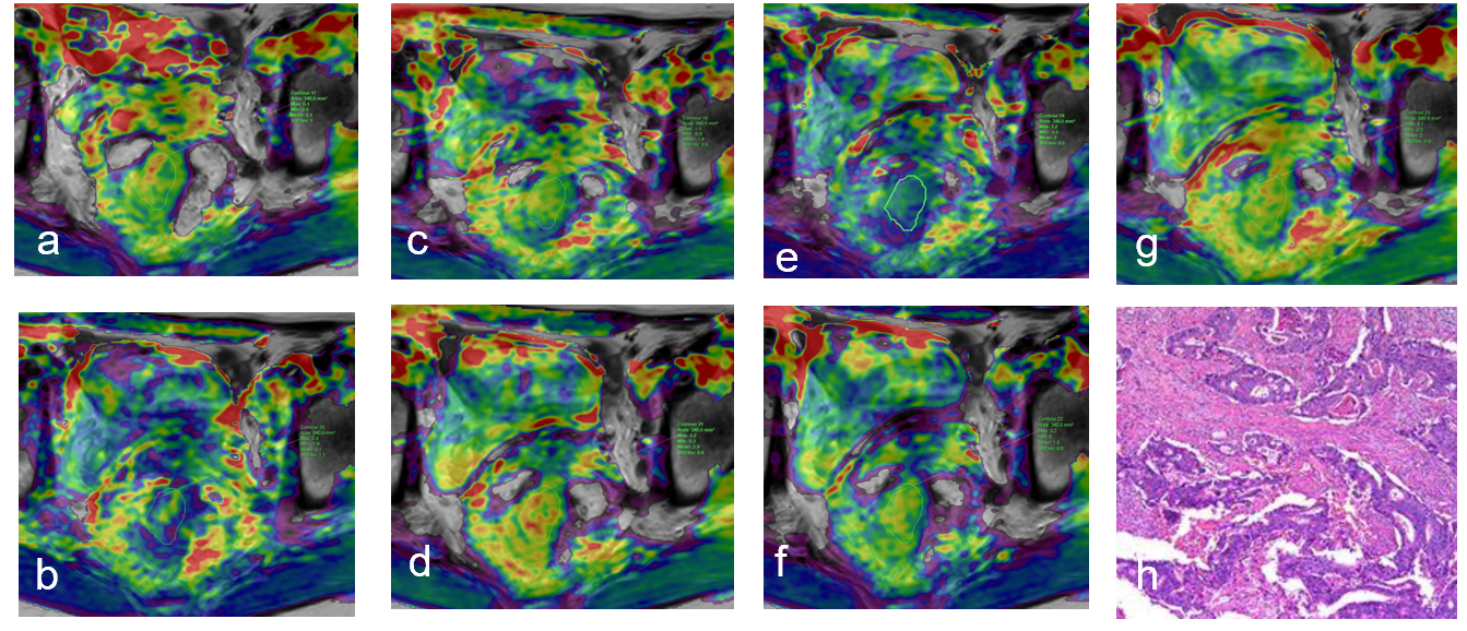
Figure1:Power level of saturation pulse and duration of saturation pulse:(a)2μT and 2 seconds (b)1.5μT and 1 second (c)1μT and 1.5 seconds (d)1μT and 1 second (e)2μT and 1.5 seconds (f)1.5μT and 2 seconds (g)1.5μT and 1.5 seconds(h)the corresponding pathology result. The image in (e) got the best imaging quality.