2436
Comparative study of APTw quantitative imaging in normal intestinal wall and rectal cancer1The first affiliated hospital of dalian medical university, DaLian, China, 2Philips Healthcare, China, Beijing, China
Synopsis
The purpose of this study was to investigate the feasibility of using APTw for the diagnosis of rectal cancer.
Introduction
Rectal cancer is the third most common cancer and the second leading cause of cancer-related death worldwide[1].Amide proton transfer-weighted imaging is one subset of the endogenous chemical exchange saturation transfer (CEST) imaging and magnetization transfer (MT) techniques. It can assess the changes in the intracellular protein concentration and PH by detecting the proton exchange between the amide protons in endogenous mobile proteins and bulk-water protons.Amide proton transfer-weighted (APTw) imaging is well applied in head、breast and recta ldiseases[2-4]. The purpose of this study was to investigate the feasibility of using APT for the diagnosis of rectal cancer.Methods
Seventeen patients with rectal cancer confirmed by postoperative pathology and fourteen volunteers were enrolled in this retrospective study. All subjects underwent 3.0T MR scan (Ingenia 3.0T CX; Philips Healthcare, Best, the Netherlands) with a 16-channel abdominal array coil. Scanning sequences: axial T2-weighted imaging (T2WI), 3D-amide proton transfer weighted imaging (3D-APT) and diffusion weighted imaging (DWI). The scan parameters were: T2WI, TR/TE= 4900/85ms, FOV=240 x 240 x 119mm3, voxel size 0.7x0.7x4mm3, scan time=2min48s.APT, TR/TE= 6500/8ms, FOV=130 x100 x49mm3, voxel size 2.0x2.0 x7mm3, scan time=3min3 s. DWI:TR/TE= 3900/60ms, FOV=240x240x119mm3, voxel size 3.0x3.0x4mm3, scan time=1min46s. All data were transferred to the IntelliSpace Portal, (Philips Healthcare). Two physicians with 3 years and 6 years experience with MRI diagnosis were employed to draw3 ROIs respectively in the signal homogenization area of rectal cancer wall on the DWI imaging and normal intestinal wall on the T2WI imaging, then getting APTw fusion graph and APTw value(Figure 1). The ROI value of the senior physician was taken as statistical analysis. The average ROI for 3 times was taken as statistical analysis. Intra-group correlation coefficient (ICC) was used to test the consistency of the measurement results of the two observers. The difference of the parameters between the two groups, the data which obeys normal distribution were performed by independent- samples T test, otherwise by Mann-Whitney test. Finally, independent-sample t test was used to test the difference between them. The ROC curve was applied to evaluate effectiveness of statistically significant parameters in the differential diagnosis of two groups.Results
The consistency of data measured by two observers was good (ICC>0.75)( Table 1).APTw value of rectal cancer was higher than that of normal intestinal wall group, their values were2.71±0.68% vs 1.55±0.68% with a statistically significant difference(P<0.05)( Table 2). The AUC was 0.878, and the corresponding diagnostic sensitivity and specificity were 94.1% and 71.4%, respectively with the APTw = 1.83 as the cut-off value(Figure2).Discussion and Conclusion
In this study, the result showed that APTw value of rectal cancer is larger than that of volunteer group. The reason may be that rectal cancer is more protein-rich.APTw imaging may serve as a preoperative and non-invasive method for the diagnosis of rectal cancer wall.Acknowledgements
No acknowledgment found.References
[1] Siegel RL, Miller KD, Fedewa SA, et al. Colorectal cancer statistics, 2017[J]. Ca A Cancer Journal for Clinicians (2017) 67(3):104-117.
[2] Togao O, Hiwatashi A, Keupp J, et al. Scan-rescan reproducibility of parallel transmission based amide proton transfer imaging of brain tumors[J]. Magn Reson Imaging, 2015,42(5):1346-53.
[3] Dula AN, Arlinghaus LR, Dortch RD, et al. Amide proton transfer imaging of the breast at 3 T: establishing reproducibility and possible feasibility assessing chemotherapy response[J].Magn Reson Med, 2013,70(1):216-24.
[4]Nishie A, Asayama Y, Ishigami K,et al.Amide proton transfer imaging to predict tumor response to neoadjuvant chemotherapy in locally advanced rectal cancer[J].J Gastroenterol Hepatol, 2019 Jan;34(1):140-146.
Figures
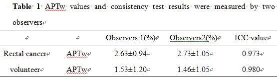
Table 1 APTw values and consistency test results were measured by two observers
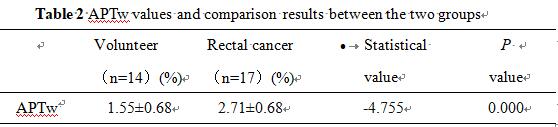
Table 2 APTw values and comparison results between the two groups
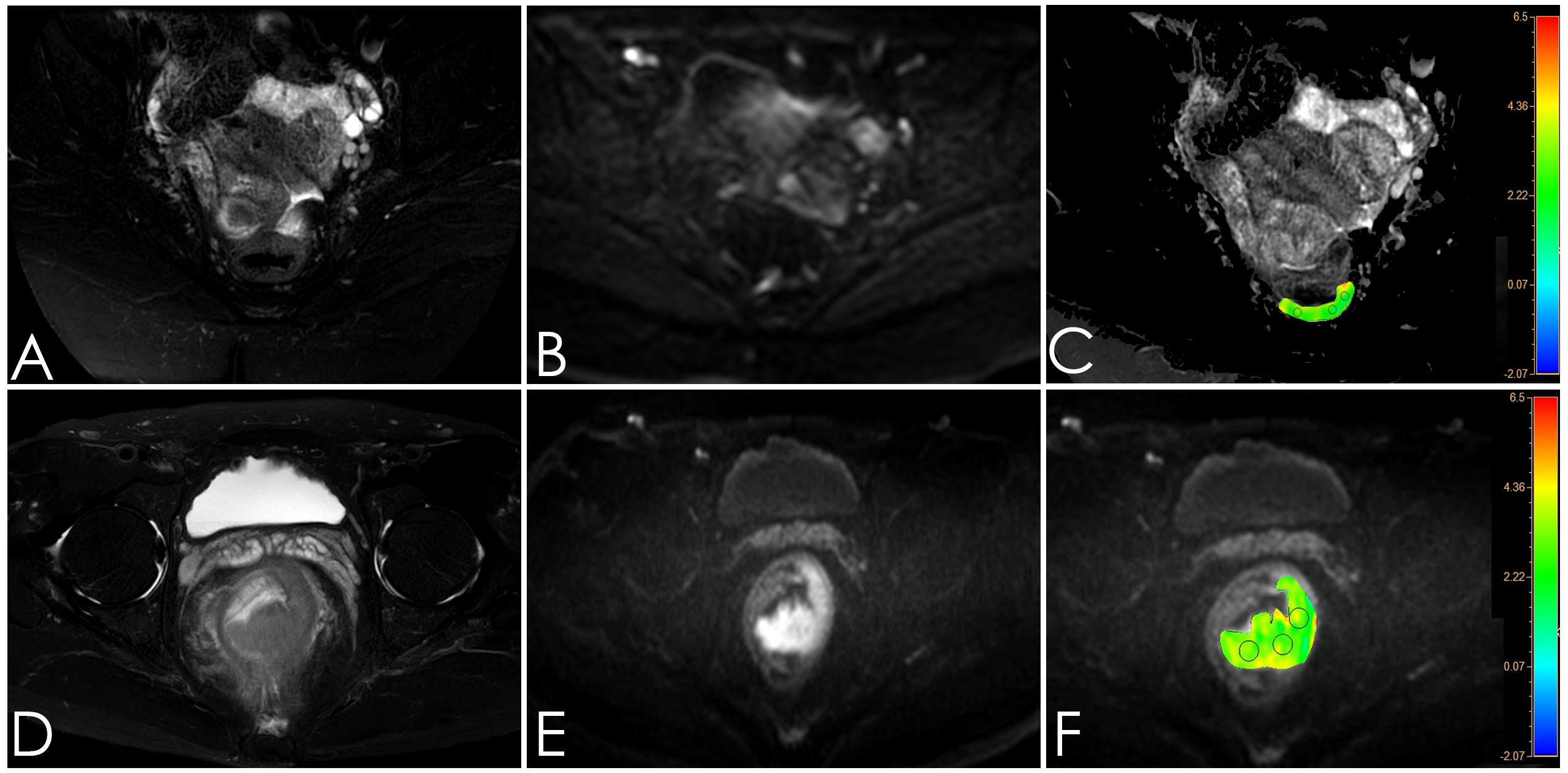
Figure 1; A-C: Female, 27 years old, normal intestinal wall,A:T2W, B:DWI, C: APTw-T2W; D-F: Male, 56 years old, rectal cancer. D:T2W, E:DWI, F: APTw-DWI. Three circular ROIs were placed on the normal intestinal wall and rectal cancer in the APTw-DWI fusion image, with the APTw values of 1.43% and 2.56%, respectively.
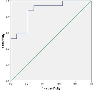
Figure2:APTw value predicted ROC curve of rectal cancer