2403
Evaluating the feasibility of predicting the multiple pathological indexes of hepatocellular carcinoma with a single MR scan1The Fourth Affiliated Hospital of Hebei Medical University, Shijiazhuang, China, 2United Imaging Healthcare, Shanghai, China
Synopsis
Imaging manifestations and pathological findings are tightly correlated to each other as they all provide the portraying of the tumor from different perspectives. We hypothesized that exploiting the quantitative markers from MR images would be a feasible way for simultaneously predicting the multiple pathological indexes. With regard to conventional “mean value” of ROI, histogram metrics, taking the tumor heterogeneity into consideration, can give full play of quantitative indicators. This research aims to preliminarily evaluate the feasibility of simultaneously predicting the multiple pathological indexes with a single Intravoxel incoherent motion (IVIM) scan by extracting multiple histogram metrics of parametric images.
Introduction
The mortality rate of HCC estimated as 8.5% and ranking 4th among all types of cancers in 2018 has been at the forefront for the long time.1 There existed multiple prognostic factors tightly related with the prognosis of HCC including the expression of AFP, Ki67, ferritin, histopathological grade, tumor size, intra-tumoral haemorrhage, capsule and so on.2 Moreover, these pathological biomarkers play significant role in assisting in making clinical decisions.3 However, there are some insurmountable disadvantages containing the invasiveness, time-consuming character and so on for accessing these pathological indexes. Nowadays, with the development of fascinating functional MRI techniques including the diffusion weighted imaging and so on, imaging approach has shown tremendous clinical potential with the advantages of non-invasive and cost-effective characters. Intravoxel incoherent motions (IVIM), able to simultaneously provide the quantification of perfusion and diffusion, has widely proven to be a powerful DWI technique for clinical application.4 However, the most widely-used quantitative metric is the mean value obtained via averaging whole ROI in the specified parametric map, which could inevitably be accompanied by the following defects: 1) the overall average severely weakens its reflection on tissue heterogeneity. 2) it’s a huge waste as there exist a lot of valuable quantitative biomarkers rather than mean value. Taking the histogram distribution of each ROI into consideration, histogram metrics act as the powerful tools for characterizing the tumor more comprehensively.5 Imaging findings and pathological results are undoubtedly the two most important means for assisting with clinical management. With the capability of characterizing tumor from different perspectives, they have proven to be tightly correlated with each other. Based on the aforementioned points, we hypothesized that there may exist significant correlations among the histogram metrics of IVIM parametric maps and multiple prognostic markers. Moreover, these correlations may render the IVIM one effective method for simultaneously providing the prediction of multiple prognostic factors, which would not only greatly ease the effort of the pathologists but also be extremely meaningful for subsequent clinical management.Methods
A total of thirty one patients diagnosed as HCC were included into this study. All patients underwent the MR examinations with a 3T MR scanner (uMR 780, United Imaging Healthcare Co Ltd). The detailed parameters of MR scanning sequences applied in this study were as the followings: IVIM sequence (TR: 4500 ms, TE: 70.0 ms, FA: 90°, thickness: 5 mm, slice gap: 1.5 mm, Matrix: 256×202, b values: 0, 10, 20, 30, 40, 60, 80, 100, 200, 400, 600, 800 s/mm2). The IVIM parametric maps including D (true diffusion Coefficent), Dp (pseudo diffusion coefficient) and f (perfusion fraction) maps were calculated according to a nonlinear bi-exponential fitting model previously reported.6 The ROI definition of all images were complemented with one abdominal radiologist with 10 years of experience. Histogram metrics were calculated with one in-house matlab based software. The pathological results including the expression of Ki67, alpha-fetoprotein (AFP) and ferritin, carcinoembryonic antigen (CA19-9), carcinoembryonic antigen(CEA) and tumor grade (Edmondson-Steiner classification) were all concluded by an experienced pathologist. Whether the patients lesions were accompanied by the intratumoral hemorrhage, capsule and fat component was also respectively assessed and determined as positive(+) (with intratumoral hemorrhage, capsule or fat component) and negative(-) (without intratumoral hemorrhage, capsule or fat component). Spearman Correlation test was performed to evaluate the correlation among the histogram metrics and pathologic indexes.Results
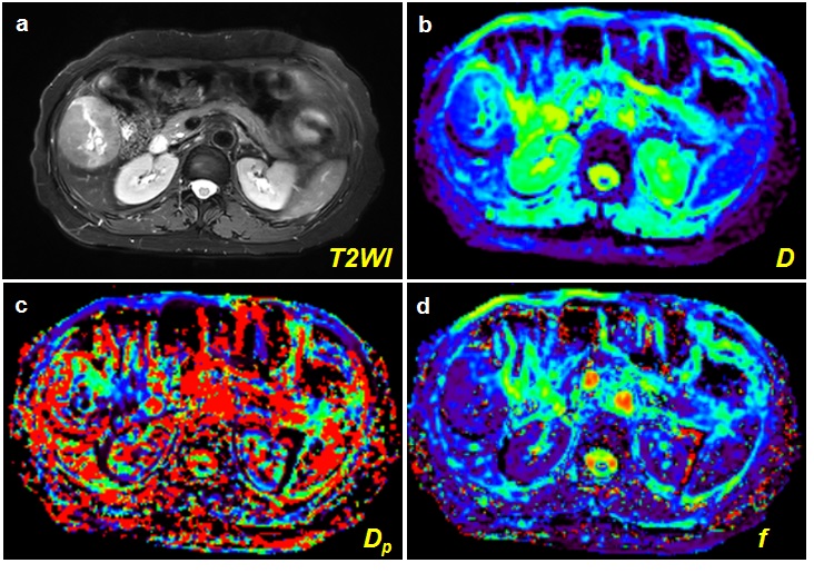
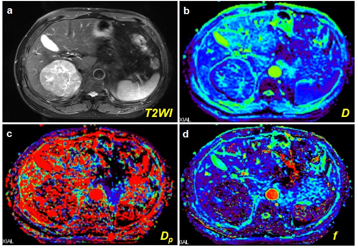
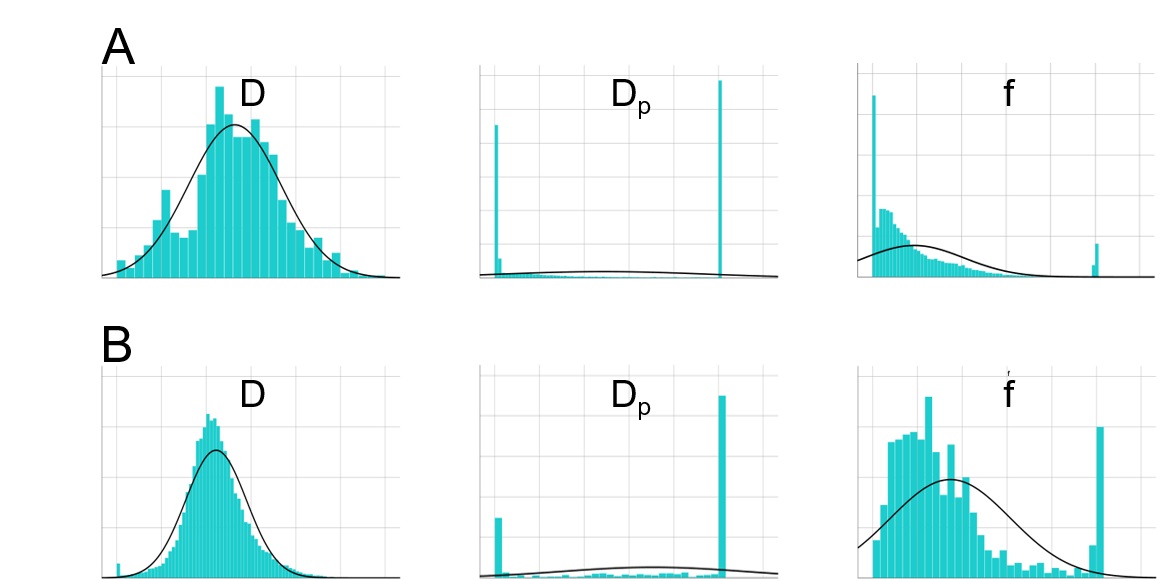
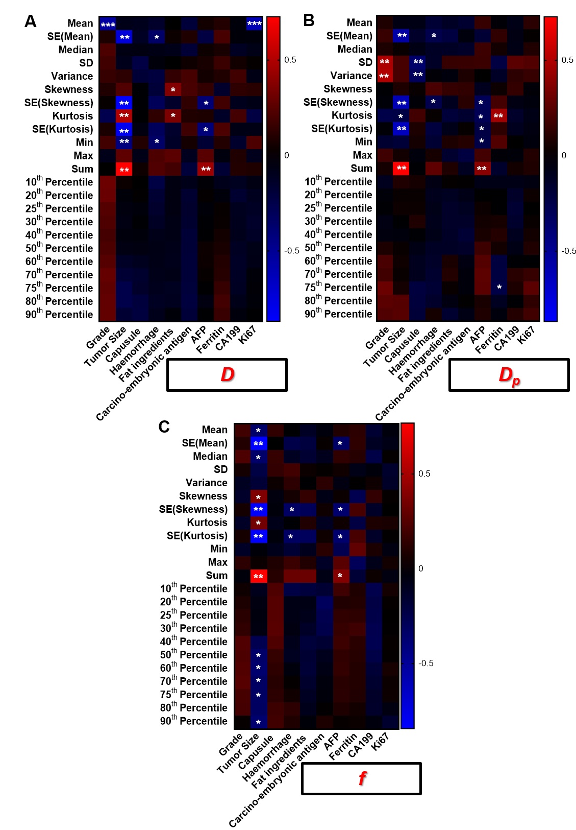
The representative MR images including T2WI images and IVIM parametric maps were displayed in the Figure 1 and Figure 2. Figure 3 displayed the distribution of IVIM histogram metrics of two HCC patients with different pathological indexes. Interestingly, although it’s hard to discover the pathological information merely by the imaging manifestations exhibited in Figure 1 and Figure 2, noticeable differences between the distributions of IVIM histogram metrics were obtained. Most importantly, as Figure 4 illustrated, numerous histogram metrics derived from IVIM parametric maps were significantly correlated with the pathological indexes (p < 0.05).Discussion
In this research, A lot of histogram metrics derived from IVIM parametric maps were significantly correlated with the pathological indexes, which was mainly caused by the following aspects: 1) As one of the most complicated biological progress, carcinogenesis is accompanied by the changes in plenty of bio-molecular signal pathways in the molecular dimension, which ultimately results in both structural and functional changes reflected by a lot of manifestations such as the variations of pathological indexes and imaging changes. Although there existed huge differences between the imaging and pathological strategy, imaging manifestations and pathological findings, in essence, are both the characterization of tumor which should be considered as a whole whose each aspects are tightly correlated. 2) Compared to the most widely-used approach in which “mean value” of ROI is applied as the quantitative index, histogram analysis is more powerful in discovering more quantitative indicators underlying the images.Conclusion
With a single MRI scan along with the extraction of histogram metrics from IVIM images, a lot of significant correlations between the metrics and pathological indexes of HCC patients could be obtained, which suggested exploiting the histogram metrcis of images would be a potential strategy for simultaneously predicting multiple pathological indexes.Acknowledgements
No AcknowledgementsReferences
1. Bray F, Ferlay J, Soerjomataram I, Siegel RL, Torre LA, Jemal A. Global cancer statistics 2018: GLOBOCAN estimates of incidence and mortality worldwide for 36 cancers in 185 countries. CA: a cancer journal for clinicians 2018;68(6):394-424.
2. Cho E, Cho HA, Jun CH, Kim HJ, Cho SB, Choi SK. A Review of Hepatocellular Carcinoma in Elderly Patients Focused on Management and Outcomes. In Vivo 2019;33(5):1411-1420.
3. Ghouri YA, Mian I, Rowe JH. Review of hepatocellular carcinoma: Epidemiology, etiology, and carcinogenesis. Journal of carcinogenesis 2017;16:1.
4. Hectors SJ, Wagner M, Besa C, et al. Intravoxel incoherent motion diffusion-weighted imaging of hepatocellular carcinoma: Is there a correlation with flow and perfusion metrics obtained with dynamic contrast-enhanced MRI? JMRI 2016;44(4):856-864.
5. Li H, Zhang J, Zheng Z, et al. Preoperative histogram analysis of intravoxel incoherent motion (IVIM) for predicting microvascular invasion in patients with single hepatocellular carcinoma. Eur J Radiol 2018;105:65-71.
6. Wei Y, Gao F, Wang M, et al. Intravoxel incoherent motion diffusion-weighted imaging for assessment of histologic grade of hepatocellular carcinoma: comparison of three methods for positioning region of interest. Eur Radiol 2019;29(2):535-544.
Figures