2389
Learning how to adapt T2 PROPELLER MR prostate imaging: going beyond PIRADS requirements with MR Deep Learning reconstruction1Clinical Research & Development, GE Healthcare, Buc, France, 2Centre Cardiologique du Nord, Saint-Denis, France, 3Global MR Applications & Workflow, GE Healthcare, Houston, TX, United States, 4Global MR Applications & Workflow, GE Healthcare, New York, NY, United States
Synopsis
To give a confident image-based prostate cancer diagnosis, PIRADS recommends using multiparametric MR (mp-MRI) exam composed of DWI and T2w sequences. By using a new deep learning-based image reconstruction algorithm, we aim to improve the utility of T2w PROPELLER images by reducing acquisition time and/or increasing spatial resolution beyond PIRADS requirements. We present quantitative analysis based on signal-to-noise ratio estimates and qualitative analysis based on delineation of anatomical structures, overall image quality, vision of thin structures.
INTRODUCTION
Demand for prostate imaging has reached unprecedented levels for prostate cancer detection, biopsy targeting, and cancer surveillance. Standardization of mp-MRI protocol in terms of spatial resolution, slice thickness and reporting described in PIRADS1 is widely adopted by radiologist.DWI is the main sequence for prostate cancer detection: it has been demonstrated to have stronger correlations with cancer grade and volume than T2w sequence. Thus, latest technical developments have been focused on DWI (multiband, reduced FOV, synthetic diffusion).
T2w sequence optimizations since PROPELLER2 have been set aside although the need for optimization remains necessary to reduce acquisition time while preserving high spatial resolution. Due to small organ size and pelvis area location, T2w PROPELLER sequence is an ideal target for improvements, especially on 1.5T MR system due to intrinsically lower signal-to-noise ratio (SNR). By increasing averages, SNR can be subsequently higher, at the expense of scan time and robustness to patient and peristaltic motions. With actual MR acquisition and reconstruction techniques, there is no acceptable way to reduce acquisition time while increasing spatial resolution beyond PIRADS recommendations.
The aim of this work is to evaluate the performance of deep learning-based image reconstruction algorithm (DLRecon PROP) to achieve higher spatial resolution without time increase.
MATERIALS AND METHOD
8 MR examinations were performed on a 1.5 T MR system (SIGNA Artist, GE Healthcare, Waukesha, WI) according to clinical standards in our institution. The study population was composed of healthy volunteers and patients referred for MR prostate exam.For patients, prostate MR exams were carried out with an axial DWI sequence (b-values: 1000 m2.s-1 and 2000 m2.s-1) and two orthogonal T2w PROPELLER sequences (axial and coronal planes). The axial PROPELLER T2w sequence parameters are FOV: 190 mm, matrix size: 340*340, slice thickness: 3mm, spacing: 0.3 mm, TE: 133ms, TR: 3400ms, bandwidth: 35.71kHz, ETL: 32, parallel imaging factor: 2 and NEX: 3.
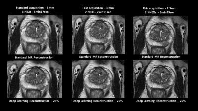
The Deep Learning (DL) reconstruction used is a deep, convolutional neural network that has been trained to improve signal-to-noise ratio and image sharpness. For DLRecon PROP evaluation, the axial T2w PROPELLER sequence was repeated first with 2 averages (Fast acquisition) and then with 2.2 mm slice thickness and adjusted averages to match standard acquisition scan time (Thin acquisition). In addition to conventional MR reconstructions, we used DLRecon PROP on all T2w PROPELLER data with different noise reduction factors (25%, 50% and 75%).
Qualitative analysis was first conducted by two blinded radiologists comparing standard and DLRecon PROP reconstructions for delineation of anatomical structures (peripheral zone, transition zone and capsule), overall image quality and sharpness. Each criterion was rated on a 5-point Likert-type scale: 5: excellent (acceptable for diagnostic use), 4: good (acceptable for diagnostic use), 3: acceptable (acceptable for diagnostic use but with minor issues), 2: poor (not acceptable for diagnostic use), or 1: unacceptable (not acceptable for diagnostic use). Quantitative analysis was performed with ROI-based SNR estimates in several regions of interest: homogenous peripheral prostate zone, fatty tissue and obturator internus muscle. An analysis of variance was used on quantitative scores to evaluate potential differences between reconstructions.
RESULTS
DLRecon PROP applied on Standard, Fast and Thin T2w Propeller acquisitions resulted in a statistically significantly higher SNR in comparison with standard reconstruction in all anatomical areas except for homogeneous peripheral zone. This result is independent from noise reduction factors used.For each exam, it has been noted that both blinded radiologists were able to distinguish DL reconstructions over standard ones due to a better image quality perception. Overall image perceived quality and delineation of anatomical structures scores were significantly higher on DL reconstruction for each sequence. However no significative difference has been found between the three noise reduction factors.
Also, by simply reducing averages, fast acquisition allows 30% scan time reduction compared with standard acquisition. DLRecon PROP allows to recover T2w PROPELLER inherent lower SNR and produces equivalent images.
Finally, for all patients with antiperistaltic injection both radiologists agreed the combination of Thin acquisition and DL reconstruction would be preferred over standard acquisition for prostate cancer staging. Indeed, radiologists noted a clear difference in image sharpness especially in peripheral zone and capsule area on patients’ exams.
DISCUSSION
Independently from sequence averages or slice thickness used, DLRecon PROP images have significantly better image quality and anatomical structures delineation. As decreasing average enable 30% scan time reduction, DLRecon PROP reconstruction provides a significant improvement of T2w clinical productivity. MR prostate sequence with reduced acquisition time provide also better exam conditions less prone to motion artifacts.Results on image sharpness were not significant due to small patient population size and will need further investigation.
CONCLUSION
This study has demonstrated the added value of a Deep Learning reconstruction in prostate T2w PROPELLER sequence leading to significant higher SNR no matter the noise reduction factor.The use of T2w PROP thicker slices sequences reconstructed by DLRecon can also be consider as promising protocol for prostate imaging allowing a better vision of details while offsetting the lack of SNR.
Acknowledgements
No acknowledgement found.References
1. Prostate Imaging Reporting and Data System, Version 2.1, 2019. American College of Radiology, 2019.
2. J. G. Pipe, « Motion correction with PROPELLER MRI: application to head motion and free-breathing cardiac imaging », Magn. Reson. Med., vol. 42, no 5, p. 963‑969, nov. 1999.
