2380
Non-contrast method for breast tumor detection using EPT based tissue conductivity maps reconstructed from routine T2-mDixon TSE images1Department of Radiology, Fortis Memorial Research Institute, Gurugram, India, 2Philips GmbH / Innovative Technologies / Research Laboratories, Hamburg, Germany, 3Philips Health Systems, Philips India Limited, Gurugram, India
Synopsis
Breast tissue conductivity map based on EPT MRI is one of the non-contrast methods that can differentiate malignant from benign breast lesions; however, the need for DCE based tumor “mask” to generate conductivity maps from the phase of VISTA images in breast challenges EPT’s role as a non-contrast method. This study demonstrates an alternative to reconstruct breast tissue conductivity maps using T2-w mDixon TSE sequence that is comparable with DCE/VISTA method, and demonstrates similarities in detecting cancerous tissues. EPT maps of breast using T2-w mDixon TSE method has the potential to help in developing non-contrast MR breast cancer screening protocol.
Introduction
Recent reports of Gadolinium deposition in tissue due to contrast injections during conventional contrast enhanced (CE) MR scans has generated increased interest among researchers to develop non-contrast alternative methods for the precision diagnosis of tumors. One such emerging non-contrast quantitative method that takes the advantage of the difference in tissue conductivities in MR is Electric Properties Tomography (EPT) methodology. In 1991, Haacke et. al. first demonstrated the possibility of derivation of electrical properties of tissues from measurable MR field which led to the estimation of tissue conductivity non-invasively using phase maps. Commonly in breast, EPT uses phase maps of a 3D-TSE (VISTA) sequence to generate conductivity maps. Clinical studies have already shown the ability of MR conductivity mapping to distinguish benign and malignant breast tumors [1-2]. However, the EPT method is challenged by the need for an ideal method of tumor delineation for conductivity reconstruction which commonly is performed by using a “mask” generated from the DCE perfusion images for breast tumors; and thus, the purpose of developing an EPT based non-contrast method for identifying malignant breast tissues fails to serve its purpose. This pilot study explores an alternative method of using the T2-weighted multi-slice mDixon TSE based tumor delineation for EPT reconstruction and compares the results with EPT reconstruction using prior knowledge of tumor using the DCE images.Methods
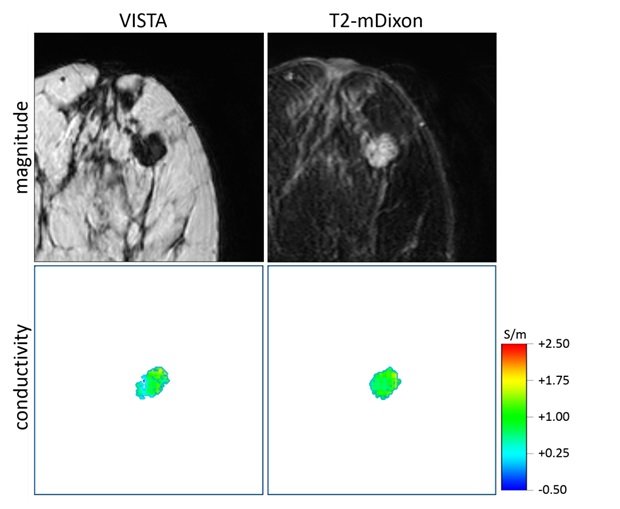
This institutional ethical committee approved study explored different EPT reconstruction methods of MR images from 11 patients with proven histopathology and BIRAD grades (2 patients with BIRAD-2, 6 patients with BIRAD-4, 2 patients with BIRAD-5 and 1 patient with BIRAD-6). Using a 7-channel breast coil at 3T MRI (Ingenia, Philips Healthcare, Best, The Netherlands) three sequences were used in this study – 1. 3D-TSE (VISTA) with phase images 2. T2 mDixon-TSE multi-slice with magnitude and phase images and 3. DCE perfusion (dynamic acquisition using 3D-TFE mDixon). Images of these three sequences were spatially registered. Binary tumor masks were derived from DCE mages. Based on the phase of the VISTA images, conductivity of each patient was reconstructed using DCE masks. Additionally, conductivity was reconstructed using the phase of the T2-weighted mDixon TSE images. In this case, no mask was used since all T2-weighted Dixon images showed sufficient tumor contrast for accurate conductivity reconstruction. After applying numerical Laplacian operator to the phase to obtain the raw conductivity, a denoising median filter was applied. The kernels for both, Laplacian and denoising filter, were shaped locally according to tissue boundaries. Conductivities were averaged first over the voxels of the individual lesions and then over each BIRADS category.Results
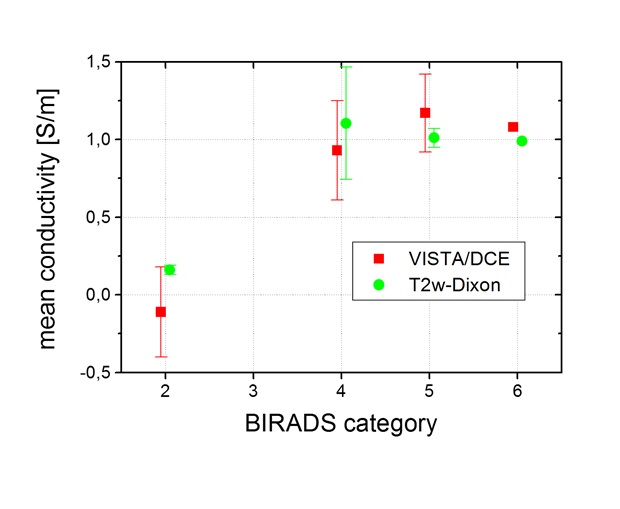
The mean lesion conductivities averaged over each BIRADS category are shown in Fig. 1. Lesions of BIRAD-2 (i.e., benign) clearly show lower conductivity values (reconstructed from VISTA phase using mask from DCE and represented as VISTA/DCE) with respect to the lesions of higher BIRAD categories (i.e., suspicious and malignant). The statistical significance of this finding is low, since only two reconstructed lesions had BIRAD-2, but agrees with findings of other studies with a higher number of patients [1,2]. On the other hand, conductivities reconstructed from T2-w mDixon TSE images confirm the finding from VISTA/DCE-based conductivity reconstructions of breast lesions.Discussion
Development of a robust non-contrast method for breast tumor detection is an unmet need especially to make MRI based routine breast cancer screening more effective. This study explores EPT as an option to address the requirement. To make EPT a truly useful non-CE method, this study adopts a new strategy to generate breast tissue conductivity maps using phase and magnitude images of T2 mDixon TSE with superior fat-suppression over conventional fat-sat techniques to bypass the commonly used tumor mask generated from breast DCE scan. The results of breast tissue conductivity of T2 mDixon TSE images are comparable with VISTA/DCE method. In addition, within a given BIRADS category, mean conductivities from T2-weighted mDixon TSE images show a trend towards a lower variability than mean conductivities from VISTA/DCE indicating possibility of achieving higher specificity using EPT for breast cancer detection. In near future, a similar study with larger patient population will be able to determine the sensitivity and specificity of the EPT maps generated from T2 mDixon TSE for detecting breast tumors to enhance the efficacy of MR based non-CE breast cancer screening.Conclusion
Breast tissue conductivity maps generated from EPT reconstruction has the potential to detect and separate benign from malignant tissues. This pilot study demonstrates that generation of conductivity maps from T2 mDixon TSE scans in a routine breast MR protocol can replace the need of DCE scans to generate breast EPT maps. This work opens up the possibility to develop EPT based non-CE breast screening protocol in future.Acknowledgements
No acknowledgement found.References
[1] Shin JW et al.; J Magn Reson Imag. 2015;42: 371;
[2] Mori N et al.,. Eur Radiol. 2019;29: 1778
Figures