2370
Feasibility of 19F Lung Imaging On A 0.5T Multi-Nuclear Upright Paramed MRI Scanner By Imaging Proton on HFC Gas.
James Harkin1, Robert Irwin1, Peter Thelwall2, and Michael Barlow1
1Respiratory Medicine, University of Nottingham, Nottingham, United Kingdom, 2Newcastle University, Newcastle upon Tyne, United Kingdom
1Respiratory Medicine, University of Nottingham, Nottingham, United Kingdom, 2Newcastle University, Newcastle upon Tyne, United Kingdom
Synopsis
Using a multinuclear 0.5T Paramed Medical Systems MROpen Upright MRI scanner the feasibility of 19F imaging was assessed. The upright and open design of the scanner makes imaging more comfortable for patients and allows for imaging in later stages of respiratory diseases. It proved to be possible to image 1H of a fluorinated gas within a single repetition and there is an expectation of at least a 2.5 fold improvement in signal when moving to 19F.
Introduction
Fluorinated gas lung imaging has the potential to revolutionise how chronic respiratory diseases are both diagnosed and their progress is monitored. After being breathed in it is possible to detect a signal in vivo from SF61, C2F62 and C3F83. Unlike hyperpolarised 129Xe/ 3He lung imaging, fluorinated gasses aren’t first hyperpolarised. Instead the short T1 of 19F in the compounds is exploited, allowing for multiple repeats within a short duration.Fluorinated gasses have the advantage over 129Xe that they don’t possess anesthetic properties and consequently be free breathed allowing for data to be obtained on wash in/ wash out kinetics4. Fluorinated compounds are also significantly cheaper and simpler to use than hyperpolarised 129Xe or 3He.Fluorinated gas MRI has previously always been performed with the volunteer horizontal in a conventional scanner. Lung function and volume, as well as arterial oxygen levels are affected by body position5. By lying supine lung function is reduced and for patients at the latter stages of chronic conditions, such as COPD, many simply can’t lay in a supine position for an extended period of time. At Nottingham the 0.5T Paramed MROpen Upright MRI scanner has two parallel TechMag Redstone spectrometers; making it the first of its kind to allow for both proton and multinuclear imaging. The subject can be imaged in a variety of positions, including supine, seated or standing. The open design is also excellent in terms of patient experience; claustrophobic reactions are virtually eliminated and the easy access and different orientations can be exploited to make the scanner better suited to disabled or paediatric cohorts.Before buying a dedicated coil tuned to 19F. The feasibility of imaging 19F was assessed for the upright scanner, through imaging the 1H of a fluorinated gas at standard temperature and pressure.Methods
An Electrolube GDP 400 air duster provided the fluorinated gas. This is cheap and contains 60-100% 1,1,1,2-Tetrafuroethane (HFC). A 10L Tedlar Bag was half filled with HFC.Neal Et. Al.’s 6 work on 19F imaging was taken as a template when working out the feasibility of imaging 19F at 0.5T. Neal Et. Al.6 imaged PFP (C3F8) at 3T with a TR of 7.5ms. The T1 of PFP in vivo is ~12.4ms. The T1 of 1H on HFC was first measured. The Clinical 3D spoiled GRE sequence from the Paramed with the minimum TE=8ms and minimum resolution (256*128*24) was modified to have a TE=2.6ms and resolution 128*32*1 (Infinite slice). The TR was then set so the ratio of T1 to TR was the same as Neal Et. Al6 and the flip angle to the Ernst Angle (57°).Results
Using two 90° RF pulses at varying TRs the drop in NMR signal was measured. The data was fitted with an R2=0.998. From this fit the T1 was calculated to be 1600ms +/-62ms with a 95% confidence interval. Using the same ratio of T1:TR as Neal Et. Al6 (TR=968ms), the half filled 10L Tedlar bag. First, with a Field of View (FOV) of 1m*1m (figure 1) then at an FOV of 0.5m*0.5m (figure 2) . A 1L bag of HFC gas was then imaged with an FOV 0.5m*0.5m (figure 3). SNRs can be seen in figure captions.Discussion
HFC can be imaged within a single scan at 0.5T and the signal is expected to improve when moving to 19F. The Bright spot in the bottom left quadrant of the images is suspected to be the hard plastics that form the imaging coil casing and is still visible when imaging an empty tedlar bag. These plastics only cause problems at short TEs due to the short T1 of 1H in the plastic. At 3T there would be expected to see 2.5 fold improvement in signal when moving to the 19F frequency with PFP from the proton frequency with HFC. 19F nuclei have a relative sensitivity of 0.84 compared to 1H. For every HFC molecule there are 2 protons and for every PFP molecule there are 8 19F atoms. However, PFP has the structure CF3-CF2-CF3 and there is chemical shift between the CF3 and CF2 resonances of 48ppm3. At 3T only the CF3 resonances are excited. However, at 0.5T the chemical shift will be 6 times smaller. This shift may be small enough to also excite the CF2 part of each molecule, making the improvement in signal even greater then 2.5 fold.Conclusions
From this preliminary work a grant submission to MRC CiC has been submitted to support the implementation of a 19F chest coil and gas delivery system. At Nottingham there is also the capability of imaging using hyperpolarised 129Xe on this scanner and there is the intention to compare both techniques in the future.Acknowledgements
Big thank you to Pete Thelwall for his expertise in fluorinated gas imaging. Also to the Haydn Green Foundation for funding my PhD.References
- Wolf, Ursula, et al. "Subsecond fluorine‐19 MRI of the lung." Magnetic Resonance in Medicine: An Official Journal of the International Society for Magnetic Resonance in Medicine 55.4 (2006): 948-951.
- Kuethe, Dean O., et al. "Imaging lungs using inert fluorinated gases." Magnetic resonance in medicine 39.1 (1998): 85-88.
- Couch, Marcus J., et al. "Inert fluorinated gas MRI: a new pulmonary imaging modality." NMR in biomedicine 27.12 (2014): 1525-1534.
- Schreiber, Wolfgang Günther, et al. "Dynamic 19F‐MRI of pulmonary ventilation using sulfur hexafluoride (SF6) gas." Magnetic Resonance in Medicine: An Official Journal of the International Society for Magnetic Resonance in Medicine 45.4 (2001): 605-613.
- Dean, Elizabeth. "Effect of body position on pulmonary function." Physical Therapy 65.5 (1985): 613-618.
- Neal, Mary A., et al. "Dynamic susceptibility contrast 19F‐MRI of inhaled perfluoropropane: a novel approach to combined pulmonary ventilation and perfusion imaging." Magnetic resonance in medicine (2019).
Figures
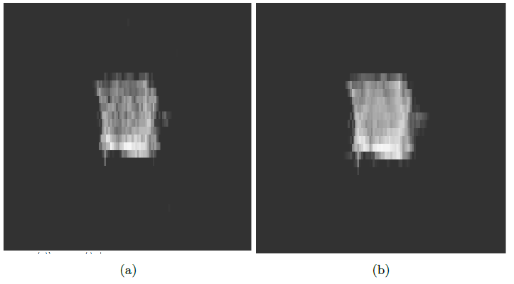
Figure 1. 1m*1m FOV: (a) 1 repetition SNR = 18.67, (b) 10 repetitions SNR= 56.76
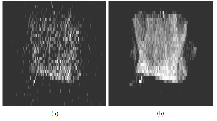
Figure 2. 0.5m*0.5m FOV: (a) 1 repetition SNR = 5.259, (b) 10 repetitions SNR = 17.89
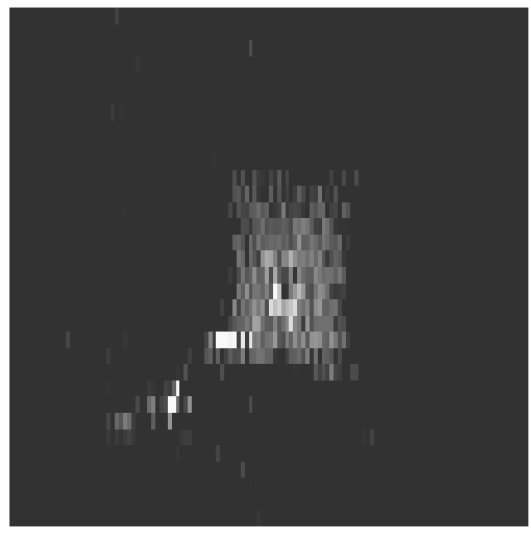
Figure 3. 0.5m*0.5m FOV 1L tedlar bag 10 repetition SNR = 8.39.