2360
Reducing Streak Artifacts in 3D Radial Imaging Using Volume-Selective Signal Acquisition1Biomedical Engineering, Sungkyunkwan University, Suwon, Korea, Republic of, 2Center for Neuroscience Imaging Research, Institute for Basic Science, Suwon, Korea, Republic of
Synopsis
The non-Cartesian sampling can obtain the image even for the number of data that does not satisfy Nyquist, but the under-sampled image is accompanied by streak artifact. Since the non-selective pulse used in conventional 3D radial UTE(C-UTE) is excite unwanted areas out of FOV, streak artifacts may appear in the image when under-sampling. Volume-Select UTE(VS-UTE) performs spin excitation using frequency-selective SINC pulses. Accordingly, the proposed method effectively suppresses the signal outside the FOV, which reduces streak artifacts. The purpose of this study is to introduce a VS-UTE sequence and to confirm that the streak artifacts are reduced effectively.
Purpose
3D radial acquisition is commonly used for lung imaging since it enables very short TE and a degree of tolerance to motion.1-2 Such a non-Cartesian sampling maintains the spatial resolution in principle even for an undersampled data but is accompanied by streak artifacts.3,4 In conventional 3D ultrashort TE (C-UTE) imaging as a representative example of 3D radial acquisition1,2, a non-selective square pulse is typically used for spin excitation with minimal TE and thus excites unwanted areas out of the region of interest (ROI). Accordingly, in lung imaging, signals from the arms, abdomen, and neck of the patient can be a strong source of streak artifacts, which is problematic especially when quantification is needed such as T1/T2 mapping, ventilation mapping etc. In this study, we suggested a method for acquiring volume-selected signals in 3D radial imaging, introducing a novel volume-selective 3D UTE sequence, dubbed VS-UTE (Volume-Selective UTE). VS-UTE was demonstrated in phantom and human lung imaging, showing significantly reduced streak artifacts which mainly originated outside the ROI.Methods
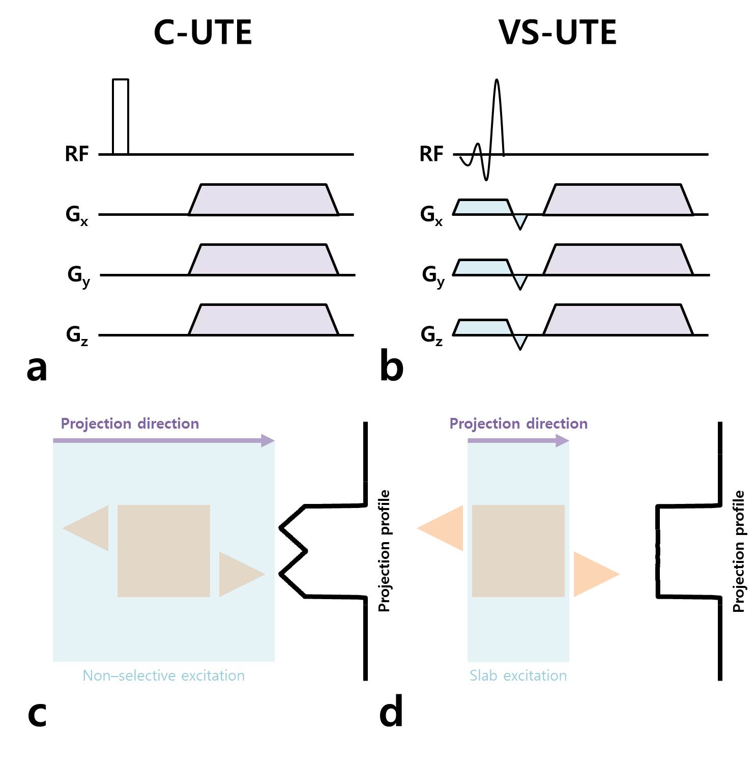
Pulse sequence description: In case of C-UTE (Fig.1a), since the entire object including the ROI is excited by a non-selective pulse depending on the coil sensitivity, the projection data contains all the information from the entire object and, as a result, streak artifacts appear due to the signal contribution from the unwanted regions such as arms, abdomen, and neck (Fig.1c). In contrast, VS-UTE performs spin excitation using a frequency-selective sinc pulse and, in particular, the readout gradient is applied perpendicular to the slab-selective gradient for each projection in order to make the projection data include only the selected information from the ROI only (Fig.1d).Experiments: VS-UTE was implemented and tested on a clinical 3T MAGNETOM Prisma scanner in comparison with C-UTE. A 34-element chest coil for signal reception was used in combination with a transmit body coil. The ROI was assumed to be same as the field-of-view (FOV).
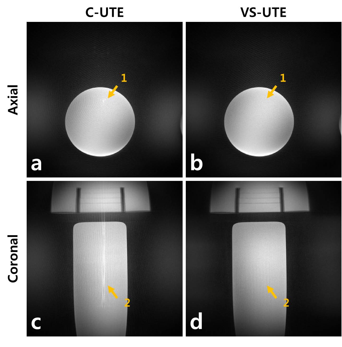
Phantom imaging: An ACR phantom, two Siemens 1900 ml saline water bottle phantoms (model #: 8624186) and a Siemens cylindrical water phantom 5300 ml (model #: 10606530) were positioned on the location of the neck, both arms, and body, respectively. FOV was set not to include the two phantoms located at both arm positions. Scan parameters were: TR = 3 ms, FOV = 300 mm3, spatial resolution = 1 mm3, flip angle = 5°, number of projection views = 60k, Pulse duration (UTE/VS-UTE) = 20/100 us, TE (UTE/ VS-UTE) = 70/200 us.
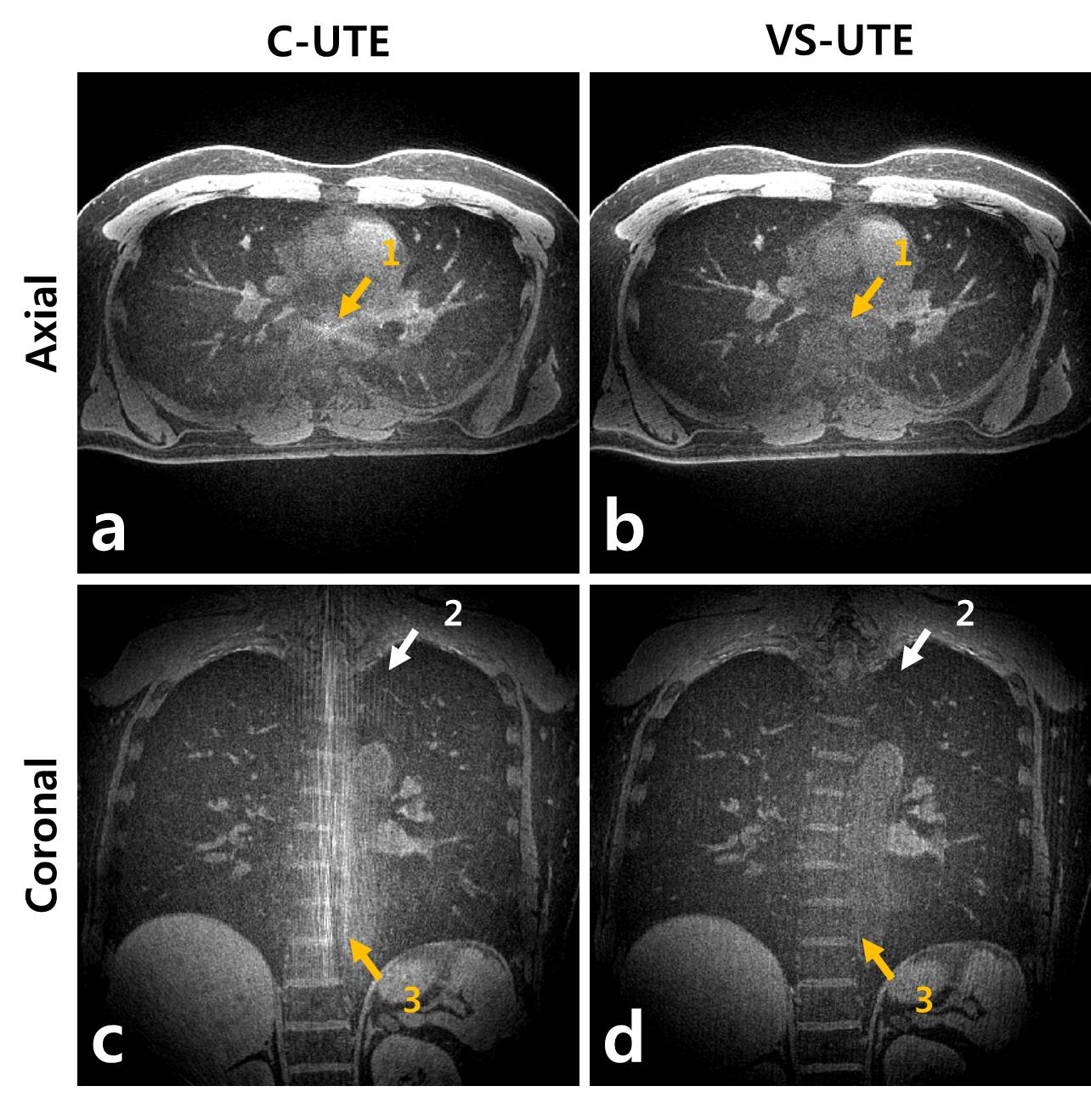
Human lung imaging: A healthy volunteer was scanned using UTE and VS-UTE in free breathing. This study protocol was approved by Sungkyunkwan University IRB and informed written consent was obtained from a healthy volunteer. A retrospective respiratory gating was performed using a self-navigation method developed by our group5 and the respiratory gating efficiency was 45%. The FOV was set not to include both arms of the volunteer, which was expected to prevent streak artifacts originated from them. A preparation spectral pulse was applied to suppress fat signals. Scan parameters were: TR = 3 ms, FOV = 320 mm3, spatial resolution = 1 mm3, flip angle = 5°, number of projection views = 130k, Pulse duration (C-UTE/ VS-UTE) = 20/100 us, TE (C-UTE/VS-UTE) = 70/200 us.
Results and Discussion
Figure 2 shows the axial (Figs. 2a, b) and coronal (Figs. 2c, d) phantom images from C-UTE (Figs. 2a, c) and VS-UTE (Figs. 2b, d). While the ACR phantom played a role as the neck, the cylindrical phantom shown in Fig.2 corresponded to the body in human chest imaging. Streak artifacts were clearly seen in both axial (like ringing artifact) and coronal slices in C-UTE (orange arrows 1, 2) as they used to appear due to the signals from the regions outside the FOV, e.g., the neck and the abdomen. In contrast, they were barely seen in VS-UTE because it acquired signals only from the inside of FOV (orange arrows 1, 2).Figure 3 shows representative axial (Fig. 3a, c) and coronal (Fig. 3b, d) slices of the healthy volunteer acquired using C-UTE and VS-UTE. In C-UTE images, streak artifacts occurred because of the incoming signals from the neck and abdomen outside the FOV (arrows 1, 2, 3), whereas the streak artifacts were suppressed in VS-UTE images because the signals outside of the FOV were effectively avoided using the volume-selective signal acquisition.
Conclusion
Here we suggested an effective way of suppressing streak artifacts in 3D radial sampling, that is, the volume-selective signal acquisition using the slab selection in the projection direction. We also proposed a new 3D UTE sequence with volume-selective signal acquisition (Volume-Selective UTE, VS-UTE), demonstrating its performance in phantom and human lung imaging. When compared to the conventional UTE imaging using non-selective spin excitation, VS-UTE significantly improved image quality with little streak artifacts by effectively suppressing the signals outside the FOV. It needs to be noted that use of a frequency-selective sinc pulse for slab selection causes a slight increase of TE in VS-UTE (~200 ms) when compared to conventional UTE (~70 ms).Acknowledgements
This work was supported by NRF-2017R1A2B2004944.References
1. Bergin CJ, Pauly JM, Macovski A. Lung parenchyma: Projection reconstruction MR imaging. Radiology 1991;179:777-781.
2. Glover GH, Pauly JM. Projection Reconstruction Techniques for Reduction of Motion Effects in MRI. Magnetic Resonance in Medicine 1992;28:275–89.
3. Du J, Thornton FJ, Fain SB, Korosec FR, Browning F, Grist TM, Mistretta CA. Artifact reduction in undersampled projection reconstruction MRI of the peripheral vessels using selective excitation. Magn Reson Med 2004;51(5):1071-1076.
4. Macdonald J, Wieben O, Nagel SK, and Johnson KM. Artifact reduction in 3D radial imaging with out-of-volume saturation pulses. In Proceedings of the 24th Annual Meeting of ISMRM, Singapore, 2016. p. 1831.
5. Park, J., Lee, S., Shin, T., Oh, S. H., & Park, J. Y. (2018). A Robust Self-navigation for Respiratory Gating in 3D Radial Ultrashort Echo-time Lung MRI using Concurrent Dephasing and Excitation. Journal of the Korean Physical Society, 73(1), 138-144.
Figures