2331
Diffusion-weighted imaging using multiplexed sensitivity encoding (MUSE) in breast cancer: comparison with single-shot echo-planar imaging1Department of Radiology, University of Cambridge, Cambridge, United Kingdom, 2Department of Radiology, Addenbrooke's Hospital, Cambridge, United Kingdom, 3GE Healthcare, Buckinghamshire, United Kingdom
Synopsis
Diffusion-weighted images acquired using single-shot echo-planar imaging (ss-EPI) often have a low resolution and are limited by blurring and distortion. This study investigated the use of MUSE (multiplexed sensitivity encoding), a multi-shot segmented-EPI technique, in the detection and characterisation of breast cancer. MUSE showed an improvement in image quality and an increased separation of malignant from benign lesions using the normalised apparent diffusion coefficient (ADC).
Introduction
Diffusion-weighted imaging (DWI) is increasingly used in the detection and characterisation of breast cancer1. Single-shot echo-planar imaging (ss-EPI) is typically used to perform DWI, however images suffer from geometric distortion and blurring due to T2* decay during readout. The necessarily lower spatial resolution of ss-EPI limits accurate measurement of the apparent diffusion coefficient (ADC) due to averaging with adjacent normal fibroglandular tissue. MUSE (multiplexed sensitivity encoding) is a segmented-EPI technique in which k-space is acquired in a number of ‘shots’ with an interleaved trajectory, and has been used in the brain to produce DWI with a higher spatial resolution and improved image quality compared to ss-EPI2. While MUSE has not yet been investigated in the breast, a number of studies have shown an improvement in image quality and diagnostic performance using RESOLVE, an alternative readout-segmented EPI technique3,4.In this study, the image quality and ADC values measured using ss-EPI and MUSE were compared. The Crété-Roffet metric5, previously adapted from the field of computer vision6, and the Mattes mutual information (MI) metric7 were used to evaluate blurring and distortion, respectively.
Methods
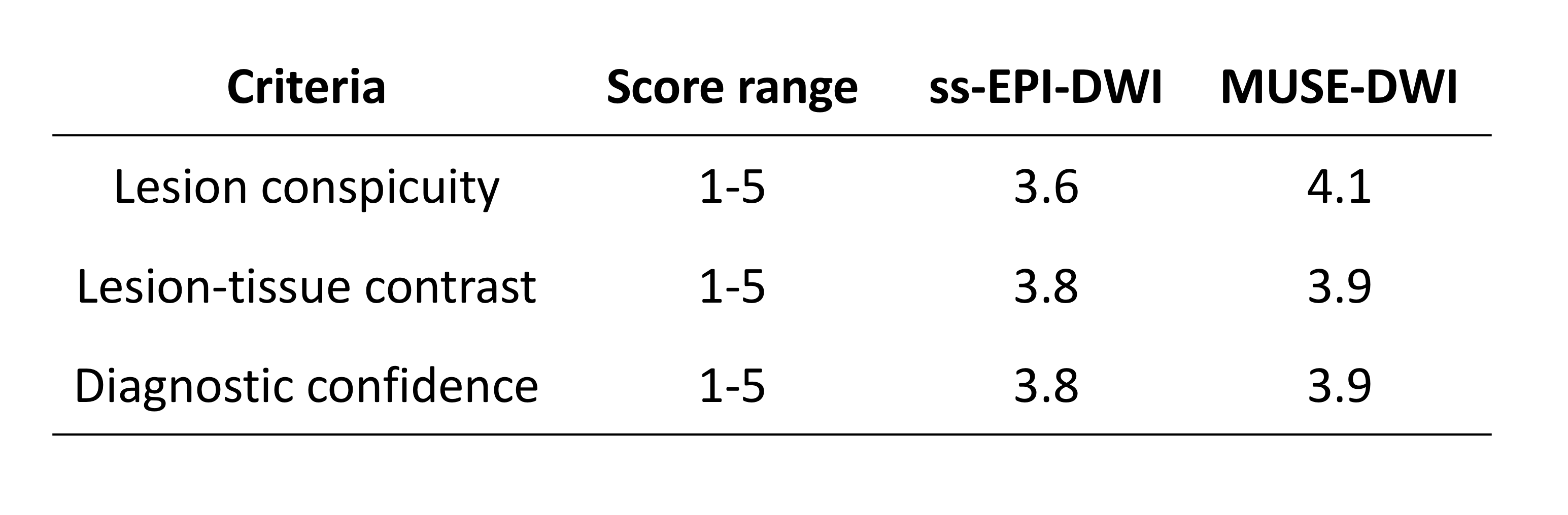
15 women with pathologically confirmed breast cancer were imaged as part of a prospective study approved by our local review and ethics boards. Imaging was performed on a 1.5T system (MR 450W, GE Healthcare, Waukesha, WI) using an 8-channel breast coil. ss-EPI-DWI and MUSE-DWI were performed. High resolution T1W dynamic contrast enhanced (DCE) images were also acquired. Scan parameters are given in Table 1.Both ss-EPI and MUSE images were scored on three qualitative image criteria: lesion conspicuity (1-5), contrast between lesion and tissue (1-5) and diagnostic confidence (1-5). Regions of interest (ROIs) were drawn in Osirix (version 8.0.1, www.osirix-viewer.com) around the whole tumour volume by a breast radiologist with reference to DCE images. Each ROI was copied onto contralateral fibroglandular tissue.
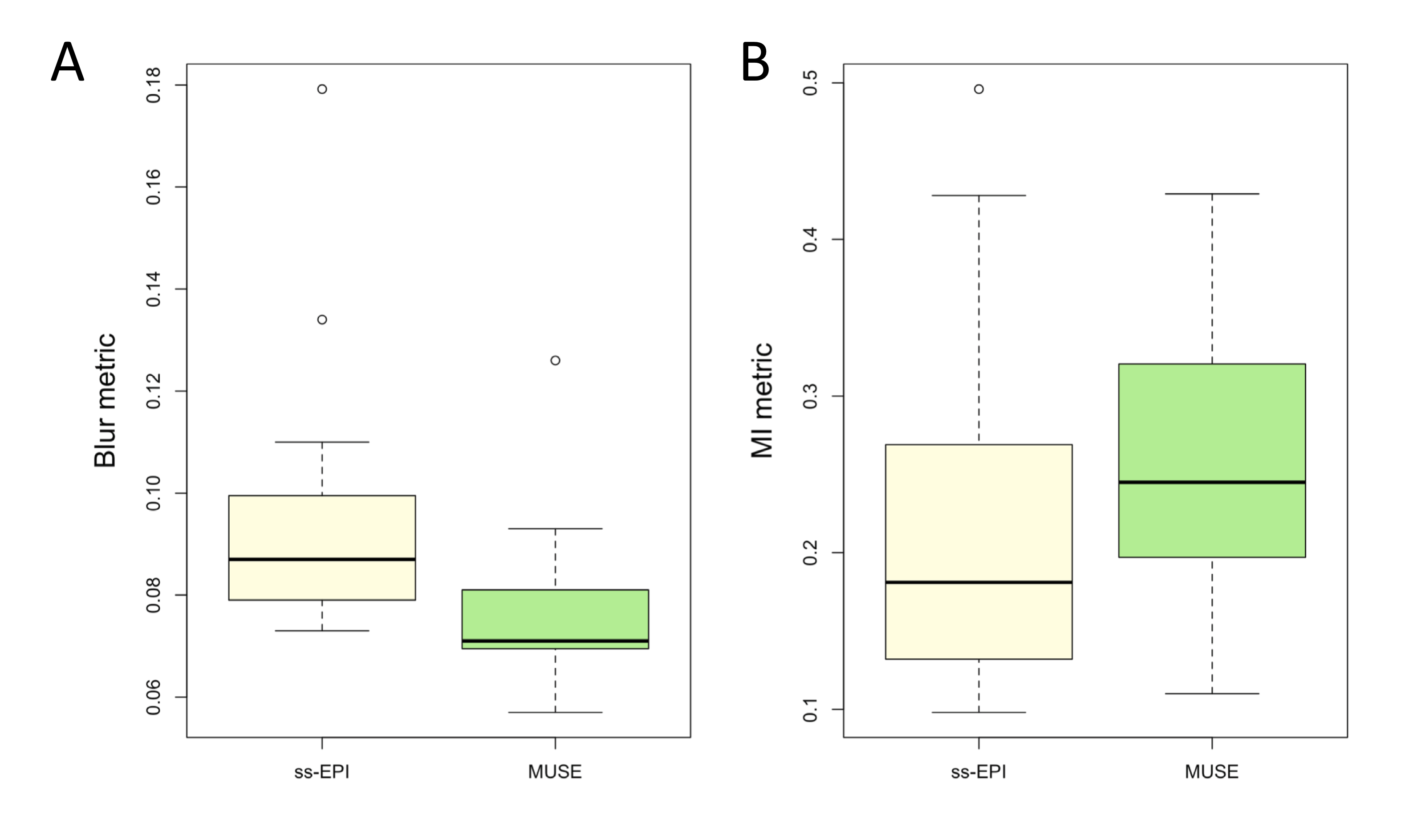
The Crété-Roffet blur metric was calculated for all ss-EPI and MUSE b = 800 s/mm2 images. Values of the blur metric range from 0 (sharp) to 1 (blurry). The MI distortion metric was calculated between ss-EPI and MUSE b = 800 s/mm2 images and the corresponding pre-contrast DCE images, resampled to the same matrix size as the DWI images. Values of the MI metric range from 0 (distorted) to 1 (not distorted). Differences in blur and distortion metrics were analysed using a Wilcoxon signed-rank test. ADC maps were generated using in-house software developed in MATLAB (version 2018b). The mean ADC of each tumour and the corresponding fibroglandular tissue ROI were measured. To account for the difference in acquired voxel size, a normalised ADC (ADCtumour/ADCfibroglandular tissue) was calculated. Differences in normalised ADC measured using ss-EPI and MUSE were compared using a paired t-test.
Results
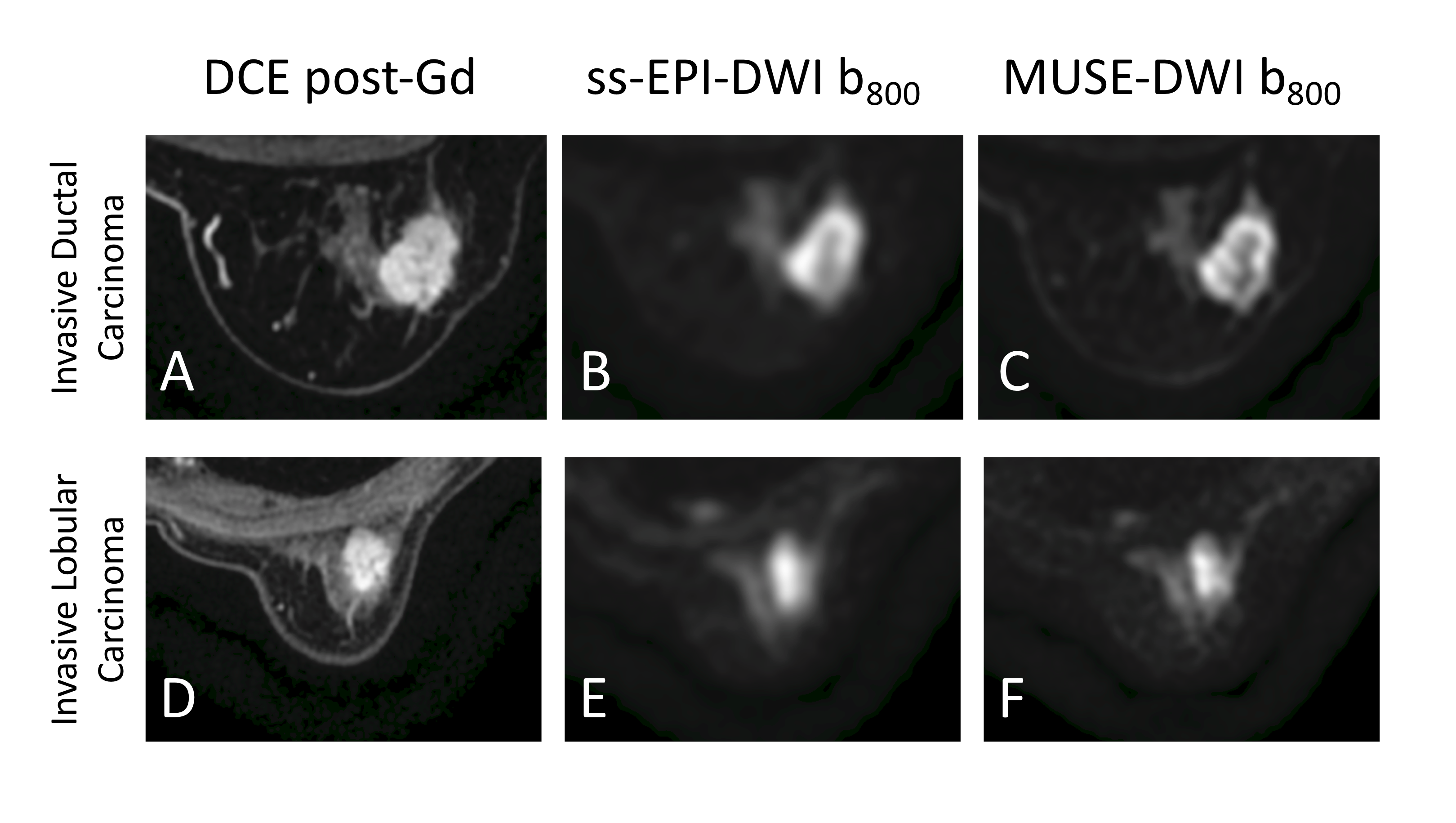
16 malignant lesions (median size 16.5mm, range 10 - 56mm) and 3 benign lesions (median size 11mm, range 10 - 16 mm) were identified in 15 patients. Figure 1 shows a comparison of image quality for an invasive ductal carcinoma and an invasive lobular carcinoma. Results of qualitative comparisons are given in Table 2. MUSE-DWI was superior to ss-EPI-DWI in all criteria. The distributions of blur and distortion metrics using ss-EPI and MUSE are shown in Figure 2. The Crété-Roffet blur metric was significantly lower for MUSE-DWI than for ss-EPI-DWI (p < 0.001), indicating less blurring. The MI metric was significantly higher for MUSE-DWI than for ss-EPI-DWI (p = 0.01), indicating more similarity to DCE images and therefore less distortion. The distributions of normalised ADC values measured using ss-EPI and MUSE are shown in Figure 3. There was no significant difference in normalised ADC values of malignant and benign lesions measured using ss-EPI or MUSE (p = 0.06 and 0.28, respectively). The separation of the mean normalised ADC values for malignant and benign lesions was greater using MUSE (0.30) than using ss-EPI (0.18).Discussion
This study demonstrated that the quality of DWI can be improved using MUSE with significantly reduced blurring and distortion. While analysis of diagnostic performance was not possible due to the low number of lesions included, the increased separation of malignant and benign normalised ADC values indicates that MUSE may be better able to characterise breast lesions than ss-EPI, improving the clinical utility of DWI. Further development aims to reduce the acquisition time of MUSE-DWI.Conclusion
The quality of MUSE-DWI is superior to that of ss-EPI-DWI, though there is an increase in acquisition time. Additional patient recruitment is required to evaluate the diagnostic performance of MUSE-DWI.Acknowledgements
No acknowledgement found.References
1. Partridge, S. C., Nissan, N., Rahbar, H., Kitsch, A. E. & Sigmund, E. E. Diffusion-weighted breast MRI: Clinical applications and emerging techniques. J. Magn. Reson. Imaging 45, 337–355 (2017).
2. Chen, N.-K., Guidon, A., Chang, H.-C. & Song, A. W. A robust multi-shot scan strategy for high-resolution diffusion weighted MRI enabled by multiplexed sensitivity-encoding (MUSE). Neuroimage 72, 41–7 (2013).
3. Kim, Y. J. et al. Readout-segmented echo-planar imaging in diffusion-weighted mr imaging in breast cancer: comparison with single-shot echo-planar imaging in image quality. Korean J. Radiol. 15, 403–10 (2014).
4. Wisner, D. J. et al. High-resolution diffusion-weighted imaging for the separation of benign from malignant BI-RADS 4/5 lesions found on breast MRI at 3T. J. Magn. Reson. Imaging 40, 674–681 (2014).
5. Crété-Roffet, F., Dolmiere, T., Ladret, P., Nicolas, M. & Crete, F. The Blur Effect: Perception and Estimation with a New No-Reference Perceptual Blur Metric The Blur Effect: Per-ception and Estimation with a New No-Reference Perceptual Blur Metric. SPIE Electronic Imaging Symposium Conf Human Vision and Electronic Imaging The Blur Effect: Perception and Estimation with a New No-Reference Perceptual Blur Metric. (2007).
6. McKay, J. A. et al. Adaptation of a Computer Vision Blur Metric to Objectively Compare High Resolution DWI Strategies in in vivo Breast Imaging. in Proc. Intl. Soc. Mag. Reson. Med. 27 (2019).
7. Mattes, D., Haynor, D. R., Vesselle, H., Lewellen, T. K. & Eubank, W. PET-CT image registration in the chest using free-form deformations. IEEE Trans. Med. Imaging 22, 120–128 (2003).
Figures