2329
DWI or dynamic contrast-enhanced curve: a retrospective analysis of MRI-based differential diagnosis of benign and malignant breast lesions1Department of Radiology, the First Affiliated Hospital of China Medical University, Shenyang, China
Synopsis
Combined with morphological and enhanced information, DWI model is superior or equal to TIC model in differentiating benign and malignant breast lesions.
Abstract
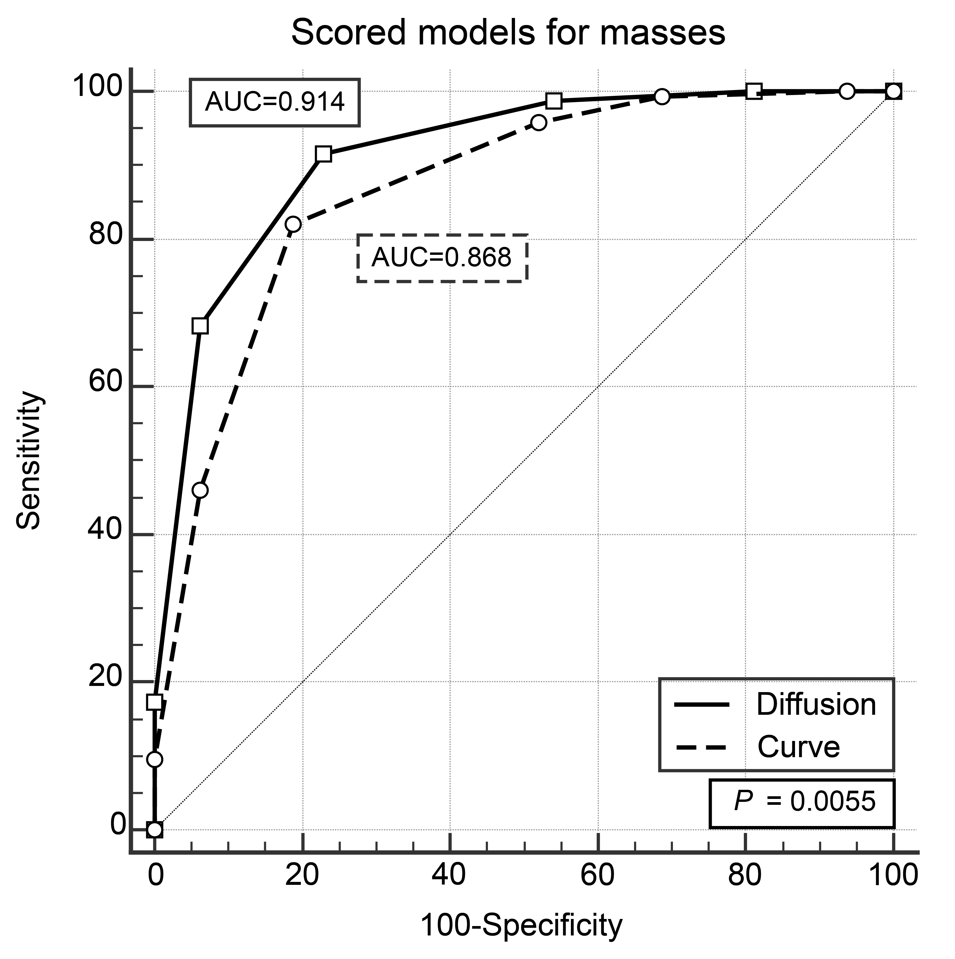
Objective To compare the diagnostic performance of models based on diffusion-weighted imaging (DWI) or time-intensity curves (TICs) in distinguishing benign and malignant breast lesions. Methods A double-blind retrospective study was conducted in 254 patients (286 suspicious lesions) who underwent dynamic contrast-enhanced magnetic resonance imaging (DCE-MRI) of the breast and obtained pathological results. Univariate binary logistic regression was applied for the apparent diffusion coefficient (ADC) value and dynamic enhancement curve (TIC) for diagnosis of benign and malignant lesions. The DWI model (ADC value + morphology + enhanced information) and the TIC model (TIC + morphology + enhanced information) were established with binary logistic regression for mass lesions and non-mass lesions respectively. The sensitivity, specificity and area under curve (AUC) were compared. The receiver operating characteristic (ROC) curves were calculated. P < 0.05 showed statistical difference. Results The sensitivities, specificities and AUC of ADC value/ TIC were 80.82%/96.80%, 77.61%/25.37% and 0.831/0.692, respectively. AUC showed significant difference between these two groups (P < 0.05). For masses, the AUC was 0.914 in DWI model and 0.868 in TIC model, with significant difference (P < 0.05). For NMEs, the AUC showed no significant difference between the DWI model and the TIC model. Conclusions Combined with morphological and enhanced information, DWI model is superior or equal to TIC model in differentiating benign and malignant breast lesions.Acknowledgements
No acknowledgement found.References
1. Medeiros LR, Duarte CS, Rosa DD et al (2011) Accuracy of magnetic resonance in suspicious breast lesions: a systematic quantitative review and meta-analysis. Breast Cancer Res Treat 126(2):273-85.
2. Orel SG, Schnall MD (2001) MR imaging of the breast for the detection, diagnosis, and staging of breast cancer. Radiology 220(1):13-30.
3. Kuhl C (2007) The current status of breast MR imaging. Part I. Choice of technique, image interpretation, diagnostic accuracy, and transfer to clinical practice. Radiology 244(2):356-78.
4. Schelfout K, Van Goethem M, Kersschot E et al (2004) Contrast-enhanced MR imaging of breast lesions and effect on treatment. Eur J Surg Oncol 30(5):501-7.
5. Abe H, Mori N, Tsuchiya K et al (2016) Kinetic analysis of benign and malignant breast lesions with ultrafast dynamic contrast‐enhanced MRI: comparison with standard kinetic assessment. AJR Am J Roentgenol 207(5):1159-1166.
6. Nogueira L, Brandao S, Matos E et al (2014) Diffusion weighted breast imaging at 3 T: preliminary experience. Clin Radiol 69(4):378-84.
7. Pereira FP, Martins G, Carvalhaes de Oliveira Rde V (2011) Diffusion magnetic resonance imaging of the breast.Magnetic resonance imaging clinics of North America 19(1):95-110.
8. Fornasa F, Pinali L, Gasparini A, Toniolli E, Montemezzi S (2011) Diffusion-weighted magnetic resonance imaging in focal breast lesions: analysis of 78 cases with pathological correlation. La Radiologiamedica 116(2):264-75.
9. Barcelo J, Vilanova JC, Albanell J et al (2009) Breast MRI: the usefulness of diffusion-weighted sequences for differentiating between benign and malignant lesions. Radiologia 51(5):469-76.
10. Parsian S, Rahbar H, Allison KH et al (2012) Nonmalignant breast lesions: ADCs of benign and high-risk subtypes assessed as false-positive at dynamic enhanced MR imaging. Radiology 265(3):696-706.
11. Nogueira L, Brandão S, Matos E et al (2014) Diffusion-weighted imaging: determination of the best pair of b-values to discriminate breast lesions. Br J Radiol 87(1039):20130807.
12. Dandan Liu, Zhaogui Ba, Xiaoli Ni, Linhong Wang, Dexin Yu, Xiangxing Ma (2018) Apparent Diffusion Coefficient to Subdivide Breast Imaging Reporting and Data System Magnetic Resonance Imaging (BI-RADS-MRI) Category 4 Lesions. Med Sci Monit 24:2180-2188.
13. D'Orsi CJ, Sickles EA, Mendelson EB et al (2013) ACR BI-RADS® Atlas, Breast Imaging Reporting and Data System— Breast MRI. 5th ed. Reston: American College of Radiology 23-144.
14. Fischer U, Kopka I, Grabbe E (1999) Breast carcinoma: effect of preoperative contrast-enhanced MR imaging on the therapeutic approach. Radiology 213(3):881-8.
15. Maltez de Almeida JR, Gomes AB, Barros TP, Fahel PE, de Seixas Rocha M (2015) Subcategorization of Suspicious Breast Lesions (BI-RADS Category 4)According to MRI Criteria: Role of Dynamic Contrast-Enhanced and Diffusion-Weighted Imaging, AJR Am J Roentgenol205(1):222-31.
16. Kuhl CK, Mielcareck P, Klaschik S et al (1999) Dynamic breast MR imaging: are signal intensity time course data useful for differential diagnosis of enhancing lesions? Radiology 211(1):101-10.
17. Schnall MD, Rosten S, Englander S, Orel SG, Nunes LW (2001) A combined architectural and kinetic interpretation model for breast MR images. AcadRadiol 8(7):591-7.
18. Schnall MD, Blume J, Bluemke DA et al (2006) Diagnostic architecturaland dynamic features at breast MR imaging: multicenter study. Radiology 238(1):42-53.
19. Dejing Ma, FengLu, Xuexue Zou et al (2017) Intravoxel incoherent motion diffusion-weighted imaging as an adjunct to dynamic contrast-enhanced MRI to improve accuracy of the differential diagnosis of benign and malignant breast lesions. MagnReson Imaging 36:175-9.
20. Bluemke DA, Gatsonis CA, Chen MH et al (2004) Magnetic resonance imaging of the breast prior to biopsy. JAMA 292(22):2735-42.
21. Kinkel K, Helbich TH, Esserman LJ et al (2000) Dynamic high-spatial-resolution MR imaging of suspicious breast lesions: diagnostic criteria and interobserver variability. AJR Am J Roentgenol 175(1):35-43.
22. Baltzer PA, Benndorf M, Dietzel M, Gajda M, Runnebaum IB, Kaiser WA (2010) False-positive findings at contrast-enhanced breast MRI: a BI-RADS descriptor study. AJR Am J Roentgenol 194(6):1658-63.
23. El Khouli RH, Macura KJ, Jacobs MA et al (2009) Dynamic contrast-enhanced MRI of the breast: quantitative method for kinetic curve type assessment. AJR Am J Roentgenol 193(4):W295-300.
24. Menezes GL, Knuttel FM, Stehouwer BL, Pijnappel RM, van den Bosch MA (2014) Magnetic resonance imaging in breast cancer: a literature review and future perspectives.World J Clin Oncol 5(2):61-70.
25. Leithner D, Wengert GJ, Helbich TH et al (2018) Clinical role of breast MRI now and going forward. Clin Radiol 73(8):700-14.
26. Thakran S, Gupta PK, Kabra V et al (2018) Characterization of breast lesion using T1-perfusion magnetic resonance imaging: Qualitative vs. quantitative analysis. Diagn Interv Imaging 99(10):633-42.
27. Igarashi T, Ashida H, Morikawa K, Motohashi K, Fukuda K (2016) Use of BI-RADS-MRI descriptors for differentiation between mucinous carcinoma and fibroadenoma. Eur J Radiol 85(6):1092-8.
28. Kuhl CK, Schild HH (2000) Dynamic image interpretation of MRI of the breast. J Magn Reson Imaging 12(6):965-74.
29. Woodhams R, Ramadan S, Stanwell P et al (2011) Diffusion-weighted imaging of the breast: principles and clinical applications. Radiographics 31(4):1059-84.
30. Pereira FP, Martins G, Figueiredo E (2009) Assessment of breast lesions with diffusion-weighted MRI: comparing the use of different b values. AJR Am J Roentgenol 193(4):1030-5.
31. Tozaki M, Maruyama K (2009) Diffusion-weighted imaging for characterizing breast lesions prior to biopsy. Magnetom Flash 2:67-70.
32. Rubesova E, Grell AS, De Maertelaer V, Metens T, Chao SL, Lemort M. (2006) Quantitative diffusion imaging in breast cancer: a clinical prospective study. J MagnReson Imaging 24(2):319-324.
33. Kul S, Cansu A, Alhan E, Dinc H, Gunes G, Reis A (2011) Contribution of diffusion weighted imaging to dynamic contrast-enhanced MRI in characterization of breast tumors. AJR Am J Roentgenol 196(1):210-7.
34. Costantini M, Belli P, Rinaldi P et al (2010) Diffusion-weighted imaging in breast cancer: relationship between apparent diffusion coefficient and tumouraggressiveness.ClinRadiol 65(12):1005-12.
35. Choudhery S, Lynch B, Sahoo S, Seiler SJ (2015) Features of non-mass enhancing lesions detected on 1.5 T breast MRI: a radiologic and pathologic analysis. Breast Disease 35(1):13-17.
36. de Almeida JR, Gomes AB, Barros TP, Fahel PE, Rocha Mde S (2016) Predictive performance of BI-RADS magnetic resonance imaging descriptors in the context of suspicious (category 4) findings. Radiol Bras 49(3):137-43.
37. Jansen SA, Fan X, Karczmar GS et al (2008) DCEMRI of breast lesions: is kinetic analysis equally effective for both mass and nonmass-like enhancement? Medical Physics 35(7):3102-9.
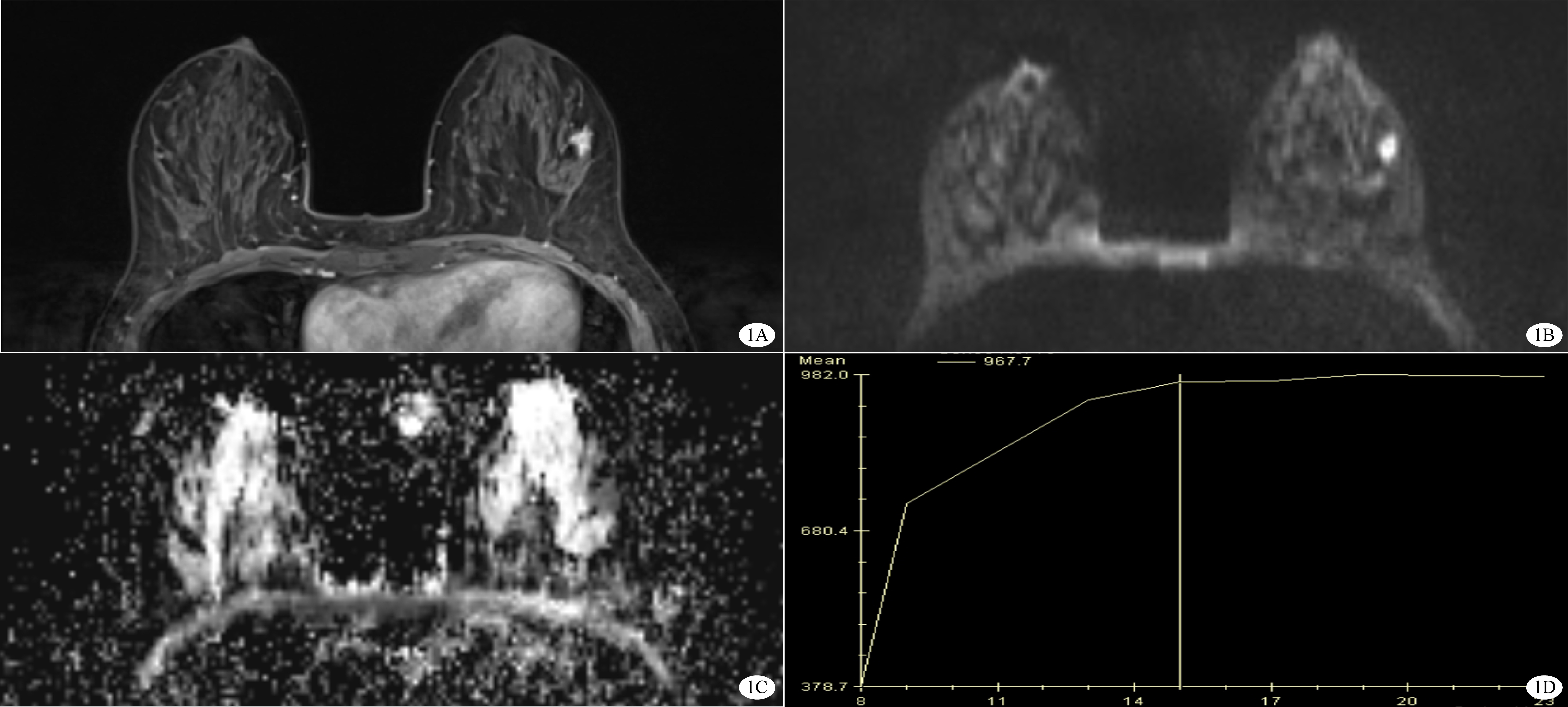
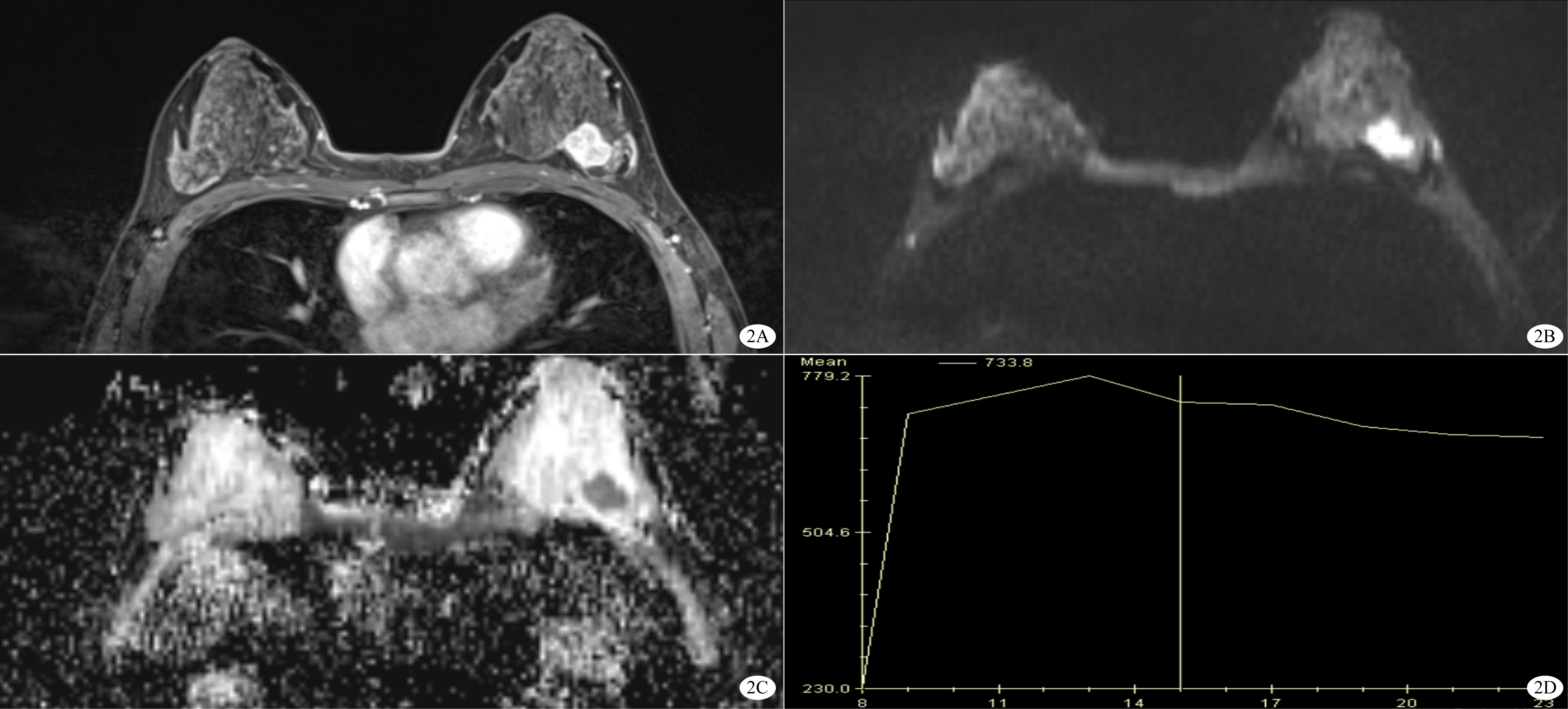
Figures