2327
Textural kinetics of suspicious breast lesions on ultrafast DCE-MRI as a lesion classifier1University of Chicago, Chicago, IL, United States
Synopsis
MRI will likely take on a greater role in breast cancer screening. However, one of the main concerns with MRI's expanded role is that it will lead to many false positives. This work aims to alleviate this problem by leveraging the advantages of ultrafast imaging of initial enhancement. Here we calculated parameters descriptive of the texture of enhancement and its changes throughout the ultrafast series. The results show that 4-D texture parameters may be useful in classifying suspicious lesions (AUC=0.75), the resulting model could have ruled out malignancy in 18% of the benign lesions analyzed, while maintaining 100% sensitivity.
Introduction
Mammography has limited sensitivity in women with dense breasts1. Recent work has shown that abbreviated MRI protocols are an effective tool for screening women with dense breasts2–4, while reducing concerns about MRI’s costs by reducing overall examination time. However, concerns about MRI’s potentially increased biopsy rates relative to mammography have been cited as a reason against its wider adoption in breast cancer screening5. Techniques that boost the diagnostic accuracy of breast MRI could help alleviate these concerns and lead to its wider adoption as a screening tool. Previous studies6–10 have reported the advantages of high-temporal-resolution (or ‘ultrafast’) DCE-MRI protocols in characterizing suspicious breast lesions via the kinetics of early enhancement. Textural analysis of breast lesions has also been shown to yield useful information for lesion classification11, although there are limited studies on texture in ultrafast imaging. 4-dimensional textural analysis (over 3 spatial and 1 temporal dimensions) yields parameters descriptive of the spatiotemporal patterns of enhancement and may yield useful information concerning tumor vascular physiology and may differentiate benign from malignant lesions12. The purpose of this study was to evaluate the performance of 4-D texture features from ultrafast DCE-MRI in differentiating suspicious breast lesions.Methods
59 patients with dense breasts (BI-RADS categories C or D) and suspicious findings on mammography (BI-RADS 4 or 5) were enrolled in this prospective study. After informed consent, patients received a research MRI prior to biopsy. Patients were scanned on 1.5T (n=5) and 3T (n=54) scanners. The DCE-MRI protocol included ultrafast imaging during the initial minute after contrast administration (0.1mM/kg gadobenic acid), with temporal resolution ranging from 2 to 10s. 4-dimensional (3 spatial and 1 temporal dimensions) gray-level co-occurrence matrices (GLCM) were calculated for rectangular ROIs encompassing each lesion through the entire ultrafast series. Twelve Haralick features13 were calculated from each lesion’s GLCM. The maximum, minimum and mean values for each of the 12 features were used to populate a logistic regression model for binary diagnosis using the biopsy results as the gold-standard. In order to eliminate unimportant parameters from the model, and to avoid overfitting, LASSO regularization was performed14. Once the most parsimonious model was identified, the classification accuracy of the model was assessed by calculating the area under the ROC curve (AUC) utilizing leave-one-out cross-validation.Results
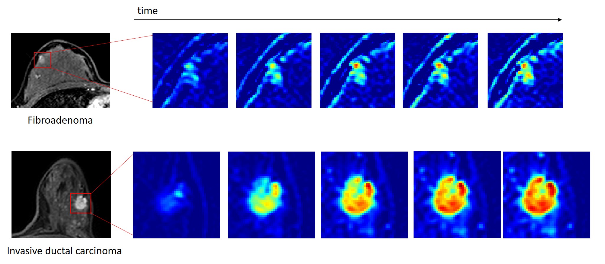
83 lesions were included in the analysis; 39 benign and 44 malignant. Representative examples of lesions imaged, and their enhancement throughout the ultrafast series, are shown in Figure 1. The logistic regression model populated with the 5 most important textural features (as identified by LASSO regularization) achieved an AUC of 0.75 (95% CI: 0.64 – 0.85). The 5 features used in the model were: maximum contrast, inertia, cluster shade, and minimum variance and sum mean. Fixing sensitivity at 100%, the model generated from the features selected could have ruled out malignancy in 7 (or 18%) of the benign lesions. This would mean an increase in the positive predictive value (PPV 3) from 53% with mammography to 58% with textural kinetic analysis of ultrafast lesions.Discussion and Conclusions
4-D texture calculated from ultrafast DCE MR images may be a useful aid in the diagnosis of suspicious breast lesions. The results suggest that the spatiotemporal patterns of very early enhancement could be a marker for malignancy. This type of analysis could help identify lesions that have a very low likelihood of being malignant, avoiding unnecessary biopsies in cases that could be safely followed-up with imaging. This analysis was performed with just one minute of post-contrast ultrafast DCE-MRI imaging. In an abbreviated MRI protocol, ultrafast imaging could be performed for the initial minute of post-contrast imaging, before switching to the high-spatial-resolution sequence, potentially boosting the PPV of abbreviated protocols.Acknowledgements
R01 CA218700, R44 CA186313, U01 CA142565References
1. Weber B, Hayes J, Phil Evans W. Breast Density and the Importance of Supplemental Screening. Curr. Breast Cancer Rep. 2018;10(2):122–130.
2. Kuhl CK, Schrading S, Strobel K, et al. Abbreviated breast Magnetic Resonance Imaging (MRI): First postcontrast subtracted images and maximum-intensity projection - A novel approach to breast cancer screening with MRI. J. Clin. Oncol. 2014;32:2304–2310.
3. Sheth D, Abe H. Abbreviated MRI and Accelerated MRI for Screening and Diagnosis of Breast Cancer. Top. Magn. Reson. Imaging. 2017;26(5):183–189.
4. Chhor CM, Mercado CL. Abbreviated MRI protocols: Wave of the future for breast cancer screening. Am. J. Roentgenol. 2017;208(2):284–289.
5. Siu AL. Screening for Breast Cancer: U.S. Preventive Services Task Force Recommendation Statement. Ann. Intern. Med. 2016;164(4):279.
6. Pineda FD, Medved M, Wang S, et al. Ultrafast Bilateral DCE-MRI of the Breast with Conventional Fourier Sampling. Acad. Radiol. 2016;23(9):1137–1144.
7. Mann RM, Mus RD, van Zelst J, et al. A Novel Approach to Contrast-Enhanced Breast Magnetic Resonance Imaging for Screening. Invest. Radiol. 2014;49(9):579–585.
8. Mus RD, Borelli C, Bult P, et al. Time to enhancement derived from ultrafast breast MRI as a novel parameter to discriminate benign from malignant breast lesions. Eur. J. Radiol. 2017;89:90–96.
9. Abe H, Mori N, Tsuchiya K, et al. Kinetic Analysis of Benign and Malignant Breast Lesions With Ultrafast Dynamic Contrast-Enhanced MRI: Comparison With Standard Kinetic Assessment. Am. J. Roentgenol. 2016;207(5):1159–1166.
10. Platel B, Mus R, Welte T, et al. Automated Characterization of Breast Lesions Imaged With an Ultrafast DCE-MR Protocol. IEEE Trans. Med. Imaging. 2014;33(2):225–232.
11. Milenkovic J, Dalmbox Drawings Light Down MU, Žgajnar J, et al. Textural analysis of early-phase spatiotemporal changes in contrast enhancement of breast lesions imaged with an ultrafast DCE-MRI protocol: Med. Phys. 2017;44(9):4652–4664.
12. Woods BJ, Clymer BD, Kurc T, et al. Malignant-lesion segmentation using 4D co-occurrence texture analysis applied to dynamic contrast-enhanced magnetic resonance breast image data. J. Magn. Reson. Imaging. 2007;25(3):495–501.
13. Haralick RM, Shanmugam K, Dinstein I. Textural Features for Image Classification. IEEE Trans. Syst. Man. Cybern. 1973;SMC-3(6):610–621.
14. Tibshirani R. Regression Shrinkage and Selection Via the Lasso. J. R. Stat. Soc. Ser. B. 1996;58(1):267–288.
Figures