2301
Motion-Corrected Pulmonary Imaging Using Spiral UTE Acquisition and Accelerated GROG-XD-GRASP Reconstruction
Eddy Solomon1, Li Feng2, Benkert Thomas 3, Moritz Schneider 4, Kai Tobias Block1, Daniel K Sodickson1, and Hersh Chandarana1
1Radiology, New York University School of Medicine, New York, NY, United States, 2Radiology, Icahn School of Medicine at Mount Sinai, New York, NY, United States, 3Siemens Healthcare GmbH, Erlangen, Germany, 4Radiology, University Hospital LMU Munich, Munich, Germany
1Radiology, New York University School of Medicine, New York, NY, United States, 2Radiology, Icahn School of Medicine at Mount Sinai, New York, NY, United States, 3Siemens Healthcare GmbH, Erlangen, Germany, 4Radiology, University Hospital LMU Munich, Munich, Germany
Synopsis
Despite recent advances in imaging techniques, respiratory motion remains a major challenge in lung MRI. This work explores ultrashort echo time (UTE) technique based on a stack-of-spirals trajectory for lung MRI. To achieve motion-resolved and high-resolution pulmonary MRI during free breathing, spiral arms were grouped into different respiratory states based on self‐navigator signals. These data were reconstructed with an eXtra-Dimensional (XD) compressed-sensing algorithm that use self-calibrating GRAPPA operator gridding (GROG). The proposed reconstruction pipeline enabled motion-compensated imaging revealing fine details of pulmonary parenchymal anatomy.
INTRODUCTION
Computed Tomography is currently the clinical gold standard for assessing lung anatomy. To avoid ionizing radiation, MRI has been proposed as an alternative modality that can provide a simultaneous evaluation of pulmonary structure and function. However, lung MRI can be challenging due to short T2* relaxation times in lung parenchyma, low proton density, and substantial respiratory motion (1). This work explores lung MRI using an ultrashort echo time (UTE) technique based on a stack-of-spirals trajectory (2,3). The sequence uses variable-duration slice encoding to minimize T2* decay, Cartesian sampling along Kz, and in-plane spiral sampling to reduce the time needed for covering k-space. To achieve motion-resolved and high-resolution pulmonary MRI during free breathing, spiral arms were grouped into different respiratory states based on self‐navigator signals (4). These data were then reconstructed with an eXtra-Dimensional (XD) compressed-sensing algorithm that used self-calibrating GRAPPA operator gridding (GROG) (5) as an initial step, to avoid repetitive gridding/regridding operations during the iterative reconstruction of the non-Cartesian data. The proposed reconstruction pipeline was tested on 5 healthy volunteers, showing improved motion-compensated imaging results as compared with conventional gridding.METHODS
All data were acquired with a work-in-progress (WIP) Spiral UTE sequence on a 3T Prisma system (Siemens Healthcare, Erlangen, Germany) using a body coil array. All human scans were performed under free-breathing conditions. Common imaging parameters included TE/TR=0.05/3.7ms, spiral duration=1600 µs, 144 slices, 2000 spiral views, no fat suppression, 1.3mm or 1.0mm isotropic resolution, and golden-angle ordering of spiral arms. The sequence uses non-selective RF excitation. Therefore, a coronal orientation was chosen to minimize the required number of through-plane phase encoding steps. Two reconstruction techniques were compared: (1) Standard gridding (NUFFT) without motion compensation, calculated directly on the scanner, and (2) the combination of GROG and XD-GRASP reconstruction (referred to as GROG-XD-GRASP and is described in the following subsection) implemented in Matlab (MathWorks, MA, USA).GROG-XD-GRASP Reconstruction: We aimed to extend the XD-GRASP approach by incorporating GROG, specifically for spiral UTE data. Using the spiral trajectory given by the machine, GROG reconstruction was integrated as a preprocessing step shifting all spiral k- space points onto a Cartesian grid. The k-space data reconstructed by GROG was then sorted according to respiratory motion data extracted from center of k-space. Next, the XD-GRASP iterative reconstruction was performed by solving the following optimization problem:
$$\arg\min_\mathbf{d} ||\mathbf{F}\mathbf{C}\mathbf{d}- \mathbf{m}||_2^2+λ||\mathbf{T}\mathbf{d}||_1$$
where $$${F}$$$ is the FFT operator, $$${C}$$$ represents coil sensitivity maps, $$${d}$$$ is the image series to be reconstructed, $$${m}$$$ is the sorted k-space data (after GROG) and $$${T}$$$ is the sparsifying transform applied along the respiratory dimension with a regularization parameter .
RESULTS AND DISCUSSION
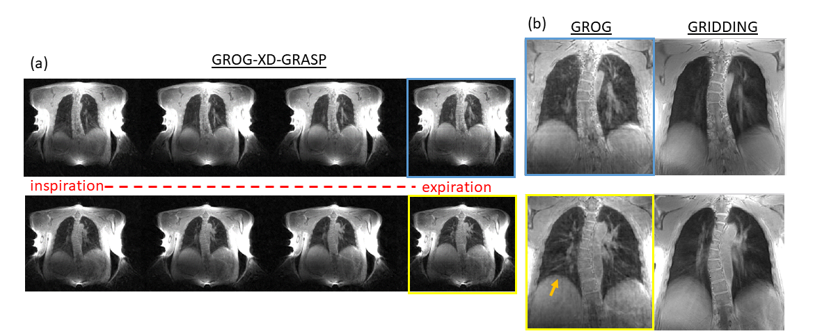
Spiral UTE data were binned into four respiratory states from inspiration to expiration (Fig.1a) based on the respiratory signal derived from the center of k-space. The subject was instructed to breath deep and steady during the scan, so that changes in lung volume could clearly be noticed in the resulting images. Next, the expiratory state was reconstructed by both the conventional gridding method and the proposed method (Fig.1b). Images generated by gridding show motion blurring (see yellow arrow) and provide less specific anatomical details. A closer comparison of these methods in another volunteer with identical spatial resolution show a clear advantage of the proposed motion-corrected reconstruction (Fig. 2, left), revealing more structures of the parenchyma. These anatomical structures can be further appreciated in the zoomed images (Fig. 2, right) of these regions. Finally, a high-resolution coronal dataset with 1.0 mm isotropic spatial resolution (Fig. 3) was binned into four respiratory states and motion‐resolved inspiration (left) and expiration (right) states were reconstructed by the two methods, demonstrating slightly finer anatomical details when using the proposed reconstruction.CONCLUSION
This work shows that self-navigation and motion-sorting enabled by spiral UTE imaging, combined with sparsity-based reconstruction, can provide additional anatomical information of potential clinical value. Moreover, when implemented with initial GROG operator to avoid repetitive gridding operations, image reconstruction can be further accelerated.Acknowledgements
We acknowledge support from NIH grant P41 EB0171813 and R01 5R01EB018308.References
- Jiang W, Ong F, Johnson KM, Nagle SK, Hope TA, Lustig M, Larson PEZ. Motion robust high resolution 3D free-breathing pulmonary MRI using dynamic 3D image self-navigator. Magn Reson Med. 2018 Jun;79(6):2954-2967.
- Qian Y and Boada FE. Acquisition-weighted stack of spirals for fast high-resolution three-dimensional ultra-short echo time MR imaging. Magn Reson Med. 2008 Jul;60(1):135-45.
- Mugler JP III, Fielden SW, Meyer CH. Altes TA, Miller GW, Stemmer A, Pfeuffer J, Kiefer B. Breath-hold UTE Lung Imaging using a Stack-of-Spirals Acquisition. Proc. Intl. Soc. Mag. Reson. Med. 23 (2015); # 1476.
- Feng L, Axel L, Chandarana H, Block KT, Sodickson DK, Otazo R. XD-GRASP: Golden-angle radial MRI with reconstruction of extra motion-state dimensions using compressed sensing. Magn Reson Med. 2016 Feb;75(2):775-88.
- Seiberlich N, Breuer F, Blaimer M, Jakob P, Griswold M. Self-calibrating GRAPPA operator gridding for radial and spiral trajectories. Magn Reson Med 2008;59:930–935.
Figures
Figure
1. (a)
Two representative lung anatomical datasets reconstructed by GROG-XD-GRASP
binned into four respiratory states using eXtra-Dimensional
(XD) self‐navigator signals. (b) The motion‐resolved GROG-XD-GRASP expiration
state (left) as compared to its counterpart slice reconstructed by conventional
gridding (right). Spatial resolution is 1.3 mm isotropic. Yellow arrow
highlights the sharper liver tip observed with GROG reconstruction.
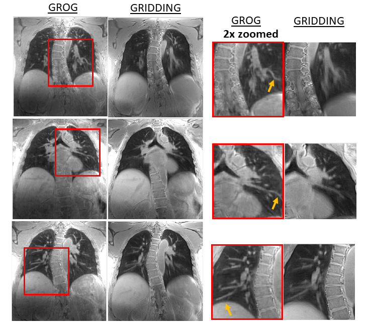
Figure 2. Three representative lung
anatomical datasets reconstructed by GROG-XD-GRASP (leftmost column) as
compared with conventional gridding (second column from the left). Spatial
resolution is 1.3 mm isotropic. The two columns on the right show zoomed
regions indicated by red rectangles.
Yellow arrows highlight lung parenchyma structures which were more
sharply defined using GROG reconstructions (second column from the right) as
compared with gridding reconstructions (rightmost column).
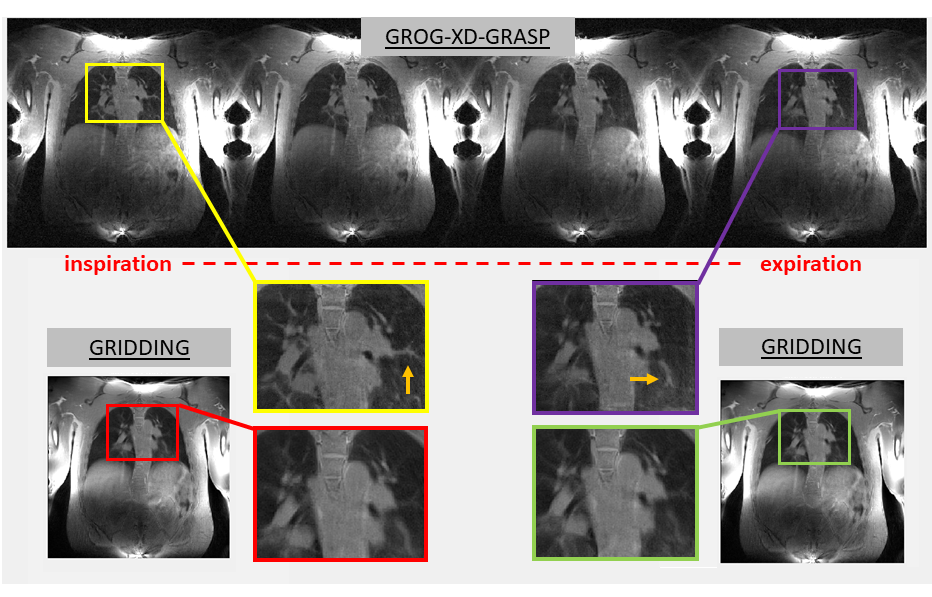
Figure 3. High resolution lung images
reconstructed by GROG-XD-GRASP (top) binned into four respiratory states.
Motion‐resolved inspiratory (top left) and expiratory (top right) states are
compared to their counterpart slices (bottom left and bottom right) reconstructed
by conventional gridding. Spatial resolution is 1.0 mm isotropic. Zoomed images
and yellow arrows at center highlight anatomical differences between the two
methods.