2296
Hyperpolarised 129Xe Imaging Using A 0.5T Upright Paramed MRI Scanner.
James Harkin1, Robert Irwin1, Joshua McAteer1, Penny Gowland2, Michael Barlow3, and Ian Hall3
1University of Nottingham, Nottingham, United Kingdom, 2Science, University of Nottingham, Nottingham, United Kingdom, 3Medicine & Health Sciences, University of Nottingham, Nottingham, United Kingdom
1University of Nottingham, Nottingham, United Kingdom, 2Science, University of Nottingham, Nottingham, United Kingdom, 3Medicine & Health Sciences, University of Nottingham, Nottingham, United Kingdom
Synopsis
Using a multinuclear 0.5T Paramed Medical Systems MROpen Upright MRI scanner, 129Xe images have been acquired within the duration of a breath hold. An active SAR monitoring system has been implemented to allow for the safe collection of data within SAR limits of chest imaging. There is the intention of applying compressed sensing to the current 129Xe sequence. The upright and open design of the scanner makes imaging more comfortable for patients and allows for imaging in later stages of respiratory diseases.
Hyperpolarised lung imaging has the potential to revolutionise how chronic respiratory diseases are both diagnosed and their progress is monitored. High resolution images can be obtained without the ionising radiation associated with CT. Images have shown great detail inside the lungs, including showing differences between healthy volunteers and patients with specific respiratory conditions1.
However, it has been shown that lung function and volume, as well as arterial oxygen levels are affected by body position2. By lying supine lung function is reduced and for patients at the latter stages of chronic conditions, such as COPD, many simply can’t lay in a supine position for an extended period of time. Even for those in the early stages, the breath hold requirements of the experiment can be difficult. Furthermore, it seems logical to extend studies to an upright orientation as the lung mechanics and function are different, yet represent a majority of time in the woken state.
At Nottingham the 0.5T Paramed Medical Systems MROpen Upright MRI scanner has two parallel TechMag Redstone spectrometers; making it the first of its kind to allow for both proton and multinuclear imaging (figure 1). The subject can be imaged in a variety of positions, including supine, seated or standing. The open design is also excellent in terms of patient experience; claustrophobic reactions are virtually eliminated and the easy access and different orientations can be exploited to make the scanner better suited to disabled or paediatric cohorts.
The scanner does have some limitations for hyperpolarized 129Xe imaging: Firstly, there is no active method of SAR monitoring; before imaging using any dedicated MNS 129Xe coils on volunteers this needed to be addressed. The Paramed also has a slow slew rate of 33Tm-1s-1 limiting the minimum TR of a sequence and when moving to a nuclear species with ~3.6 times the gyromagnetic ratio the limitations are amplified. For Lung imaging the sequence must be completed within one breath hold placing constraints on the maximum resolution.
The clinical 3D GRE proton sequence on the Paramed was first broadbanded so it functioned at the 129Xe resonant frequency in the scanner. The TR of the sequence was then reduced from 16.4ms to 13.4ms by modifying the duration, timing and amplitude of the phase encoding and spoiling gradients. The current coil used for imaging is a single loop surface coil used for both transmit and receive. When imaging, saline bags were placed on top of the coil to load it (simulating tissue around the lung) and the 129Xe phantom was placed on top of the saline bags (simulating the lung itself). Compressed sensing has also been shown in practice to be effective when undersampling up to 2.5 times the clinical 3D GRE proton sequence. This will be applied to the 129Xe sequence to allow for higher resolution scans within a breath hold.
The Paramed uses a Tomco BT02000-AlphaS 100kHz-30MHz 2kW RF amplifier. The RF Forward power can be sampled at ~50dB. The exact attenuation of the sample was measured by measuring the actual forward power as well as the output of the sample. An analogue integration circuit was then built. In the event of a prescribed power being exceeded a flag is raised which sets the transmit gate to 0 preventing any future RF input.
Using a FOV of 40cm*30cm*30cm, resolution of 256*128*8 and 6° flip angle a 1.6L tupperware phantom filled with 129Xe and with 2 voids; a glue stick and sealed glass vial was imaged in <14s (figure 2). The voids are clearly visible and the base of the glue stick which is twisted (a feature of length ~1mm) can be distinguished.
Using the same flip angle, resolution and FOV a length of tubing with internal diameter 3mm was imaged (figure 3).
The TOMCO amplifier was shown to provide 64.8 dB of gain. There was an excellent fit between the RF sample voltage and the output power with an R2=0.99 (figure 4). From the fitted curve the actual attenuation of the sample from the actual forward power was found to be 44.8dB not the ~50dB expected.
Hyperpolarised 129Xe images have been taken on phantoms using a simple surface coil and managed to distinguish features of ~1mm. The FOV and duration of the scan was chosen to allow for direct translation into humans. A 4 receive channel 129Xe body coil will imminently be arriving from Pulse Teq. Using this coil in tandem with a reliable SAR monitoring system data taken on this coil in human cohorts both supine and upright will be presented. There is the intention to apply compressed sensing to the 129Xe 3d scan to push to higher resolutions within a breath hold.
Acknowledgements
No acknowledgement found.References
- Dregely I, Mugler JP, Ruset IC, et al. Hyperpolarized Xenon-129 gas-exchange imaging of lung microstructure: first case studies in subjects with obstructive lung disease. J Magn Reson Imaging. 2011;33(5):1052-62.
- Dean, Elizabeth. "Effect of body position on pulmonary function." Physical Therapy 65.5 (1985): 613-618.
Figures
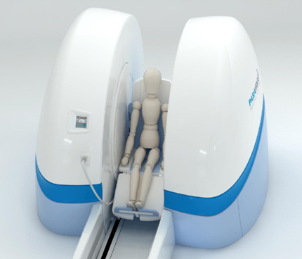
Figure1. Paramed Medical Systems MROpen Upright MRI Scanner
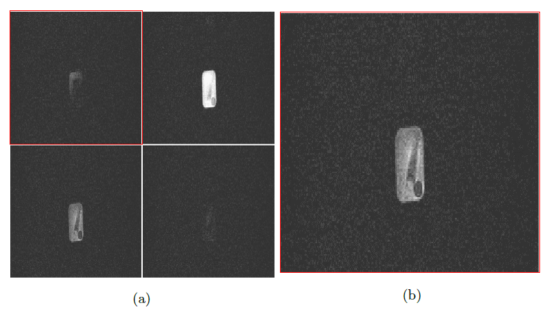
Figure 2. 8 slice 3d spoiled GRE slices of 129Xe phantom (a) Slices 4, 5, 6 and 7. (b) Slice 6, The glue stick and glass vial voids can clearly be distinguished.
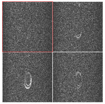
Figure 3. 8 slice 3d spoiled GRE slices of 129Xe phantom slices 3, 4, 5 and 6. 3mm internal diameter plastic tubing used as 129Xe phantom.

Figure 4. RF SAMPLE P2P voltage against the power output from the Tomco fitted to y=10^[(alog10(x) + b], R2 0.999.