2265
Comparison between 4D Flow in abdominal magnetic resonance imaging for portal vein using SENSE and Compressed SENSE
Yuli HUANG1, Li ZHU2, Haiyang DONG2, Yuan QU3, and Yan WANG3
1MR, Philips Healthcare (Suzhou) Co., Ltd, SUZHOU, China, 2Radiology, Shanghai Chest Hospital, Shanghai JiaoTong University, Shanghai, China, 3Radiology, People’s Hospital of Xinjiang Uygur Autonomous Region, Urumqi, China
1MR, Philips Healthcare (Suzhou) Co., Ltd, SUZHOU, China, 2Radiology, Shanghai Chest Hospital, Shanghai JiaoTong University, Shanghai, China, 3Radiology, People’s Hospital of Xinjiang Uygur Autonomous Region, Urumqi, China
Synopsis
4D Flow MRI is a technique that can characterize vascular structure and hemodynamics. Compared with SENSE, Compressed SENSE can further reduce scanning time using the sparse characteristics of MR images. This study aims to investigate the feasibility of 4D Flow MRI with Compressed SENSE by comparing with 4D Flow MRI with SENSE. 4D Flow dynamic angiography of hepatic portal vein using SENSE and Compressed SENSE could be completed within 5 minutes with optimized parameters. The scanning time of Compressed SENSE 4D Flow was shorter than that of SENSE 4D Flow.
INTRODUCTION
Portal hypertension is a common clinical prognosis of cirrhosis, which often leads to blood disorders, collateral circulation, and in severe cases portal vein blood flows away from the liver.1,2 4D Flow MRI is a magnetic resonance (MR) imaging technique which can characterize vascular structure and hemodynamics.3,4 However, long scanning time with 4D Flow scan limited its clinical application. Recently, Compressed SENSE (CS) has been validated as an efficient acceleration technique in fast MR imaging compared with SENSE.5 This study aims to investigate the possibility of 4D Flow MRI with CS by comparing with 4D Flow MRI with SENSE.METOHDS
Study sample: Ten female and twelve male healthy controls (mean age, 38.2 ± 13.2 years) without any contraindication to MR scanning underwent abdominal MR imaging after fasting. All the parts of this study were approved by local institutional review board and written consent form was obtained from each subject. MR imaging: Abdominal MR imaging was performed on a 1.5 Tesla MR scanner (Achieva, Philips Healthcare) with an 8-channel abdominal coil and a 12-channel spine coil. Firstly, a 2D balanced FFE (bFFE) scan was performed with breath-hold to determine the correct orientation of oblique section parallel to portal vein. Secondly, maximum flow velocity in portal vein was measured by QFLOW was set as the input of velocity encoding. Finally, 4D Flow MR imaging with SENSE and CS using ECG gating was performed with the following parameters: 3D fast field echo, number of excitations 3, repetition time 4.9 ms, echo time 3.5 ms, flip angle 10°, field of view 320 × 320 mm2, slice number 36, slice thickness 3 mm, voxel size = 3 × 3 × 3 mm3. The accelerator factor was 2 × 1.8 in acquisition with SENSE and 4 with CS. Fifteen phases were acquired in a cardiac circle. Data Analysis: Abdominal 4D Flow images were reviewed by two radiologists with >10 years’ experience blinded to each other. Each reviewer drew five regions of interest (ROIs) >50 mm2 in main portal vein (MPV), superior mesenteric vein (SMV), splenic vein (SV), left and right portal vein (LPV, RPV), respectively. Signal-to-noise ratio (SNR) was calculated in MPV, SMV, SV, LPV and RPV with the following equation:SNR=Svessel/SD(Snoise)
where Svessel represented mean value of signal in PV and its branches. The maximum flow velocity and flow volume were calculated in MPV, SMV, SV, LPV, and RPV using GTFlow (Zurich, Switzerland). Additionally, 4D Flow reconstructed streamline images were assessed by two reviewers according to a 1 to 4 rating principle which was used to evaluate the image quality (Figure 1). The scanning time of 4D Flow with SENSE and CS was recorded separately. Statistical Analysis: The SNR was compared between the ROIs in 4D Flow images acquired with SENSE and CS using a paired t test. The difference between the image quality of the reconstructed streamline was compared using a rank sum test. A p<0.05 was considered as statistically significantly and all statistical analyses were conducted with SPSS 25.0 (IBM Inc., USA).
RESULTS
There was no significant difference in SNR between the ROIs in the images acquired with SENSE and CS (P>0.05). No significant difference was found in maximum flow velocity and flow volume of SV, SMV, MPV, LPV and RPV (All P>0.05). Additionally, the image quality of streamline of SV, SMV, MPV, LPV and RPV was similar (Figure 2) and no statistically significant difference was found in image quality score (P>0.05) (Table 1). The score of image quality was consistent between the two physicians (kappa = 0.89). There was a significant difference in scanning time between 4D flow with SENSE and CS (293.70 ± 31.63s vs. 271.45 ± 39.64s, p <0.05).DISCUSSION
In the present study, 4D Flow with CS acceleration could shorten scanning time without loss of SNR in portal vein MR imaging compared to that with SENSE acceleration. 4D Flow dynamic angiography of hepatic portal vein using SENSE and CS could be completed successfully within 5 minutes with optimized parameters.6 After good preparation of abrosia and water deprivation before examination, 4D Flow scanning combined with diaphragm navigation and abdominal wall navigation technology could minimize the impact of respiratory movement on image acquisition. The image quality and reconstructed streamline tracking of the two methods were similar.7CONCLUSION
Fast 4D Flow imaging with SENSE and CS exhibited similar SNR and image quality in portal vein imaging, but acquisition with CS might be a more potential acceleration method in 4D Flow due to its shorter time consumption.Acknowledgements
No acknowledgement found.References
- Stankovic Z, Frydrychowicz A, et al. MR-based visualization and quantification of three-dimensional flow characteristics in the portal venous system. Journal of magnetic resonance imaging: JMRI. 2010; 32(2):466–475. doi: 10.1002/jmri.22248.
- Stankovic Z, Csatari Z, Deibert P, et al. Normal and altered three-dimensional portal venous hemodynamics in patients with liver cirrhosis. Radiology. 2012; 262(3):862–873. doi: 10.1148/radiol.11110127.
- A Roldán-Alzate, PhD, et al. In vivo Validation of 4D Flow MRI for Assessing the Hemodynamics of Portal Hypertension. J Magn Reson Imaging. 2013 May; 37(5): 1100–1108. doi: 10.1002/jmri.23906.
- Benjamin R Landgraf, BS, et al. Effect of Temporal Resolution on 4D Flow MRI in the Portal Circulation. J Magn Reson Imaging. 2014 April ; 39(4): 819–826. doi: 10.1002/jmri.24233.
- Tariq U, Hsiao A, Alley M, et al. Venous and arterial flow quantification are equally accurate and precise with parallel imaging compressed sensing 4D phase contrast MRI. J Magn Reson Imaging. 2013 Jun; 37(6):1419–1426. doi: 10.1002/jmri.23936.
- Roldan-Alzate A, Frydrychowicz A, Niespodzany E, et al. In vivo validation of 4D flow MRI for assessing the hemodynamics of portal hypertension. J Magn Reson Imaging 37:1100–1108. doi: 10.1002/jmri.23906.
- Buonocore MH. Visualizing blood flow patterns using streamlines, arrows, and particle paths. Magn Reson Med. 1998; 40:210–226. doi: 10.1002/mrm.1910400207.
Figures
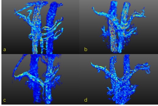
Figure 1. Quality scoring of 4D Flow streamline image of hepatic portal
vein and its branches. A, the image evaluation is 4; B. the image evaluation is
3; C. The image evaluation is 2; D, the image evaluation is 1.
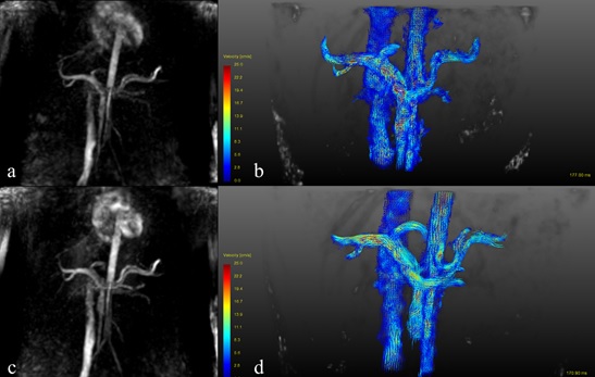
Figure 2. Image reconstruction and 3D streamline visualization of SENSE and
Compressed SENSE 4D Flow. a). Reconstructed image of SENSE 4D Flow, b) 3D
streamline visualization of SENSE 4D Flow, c). Reconstructed image of
Compressed SENSE 4D Flow, d) 3D streamline visualization of Compressed SENSE 4D
Flow. Both streamline visualization could illustrate the blood flow of portal
vein and its branches.
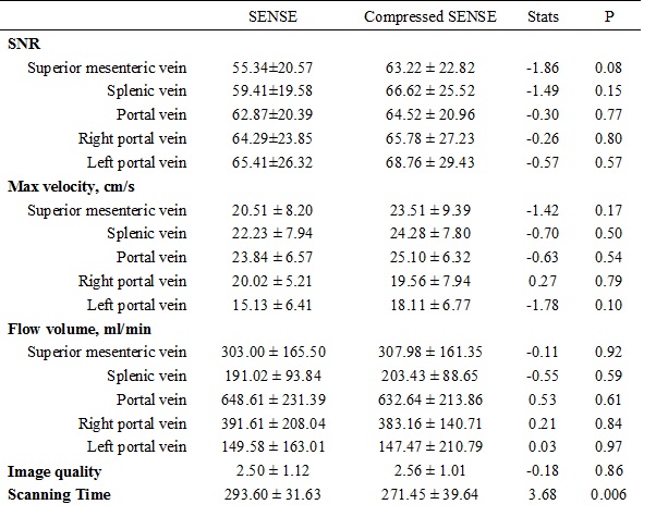
Table1.
Statistical analysis results of the SNR, maximum velocity, flow volume, image
quality, and scanning time of 4D Flow MRI with SENSE and Compressed SENSE.