2251
Slice Encoding for the Reduction of Outflow Signal Corruption in Cine Balanced Steady State Free Precession Imaging1Radiology, University of California, Los Angeles, Los Angeles, CA, United States, 2Radiology, The Unversity of Chicago, Chicago, IL, United States
Synopsis
Balanced steady-state free precession (bSSFP) is susceptible to outflowing signal because of it features no spoiling. This results in misregistrated artifacts that hamper the cardiac evaluations. We propose a method that tackles this issue in the acquisition process by spatially encoding for this outflowing signal to prevent it from projecting onto the imaged slice.
Introduction
Two-dimensional balanced steady-state free precession (2D bSSFP) is the standard sequence used in cardiac cine MRI1,2. Because of its balanced3-7 and refocused8,9 nature, this sequence achieves a high T2/T1 dependent signal-to-noise ratio (SNR). Along with inflow-enhanced contrast from through-plane flow10,11, these features make 2D bSSFP desirable for cardiac evaluations including myocardial structure, calculating blood-ejection fraction, and assessing ventricular function12-14. However, the features that make 2D bSSFP useful also pose a nuisance: the lack of spoiling provides a susceptibility to outflowing signal15-17. This effect, in combination with higher off-resonances seen at 3T, often causes outflow-induced image artifacts. In this work, we propose a through-slice phase-encoding (slice-encoding) scheme similar to SEMAC18 that expands our through-slice field of view (FOV) beyond the nominal slice-thickness to prevent outflowing signal from corrupting the intended image.Theory
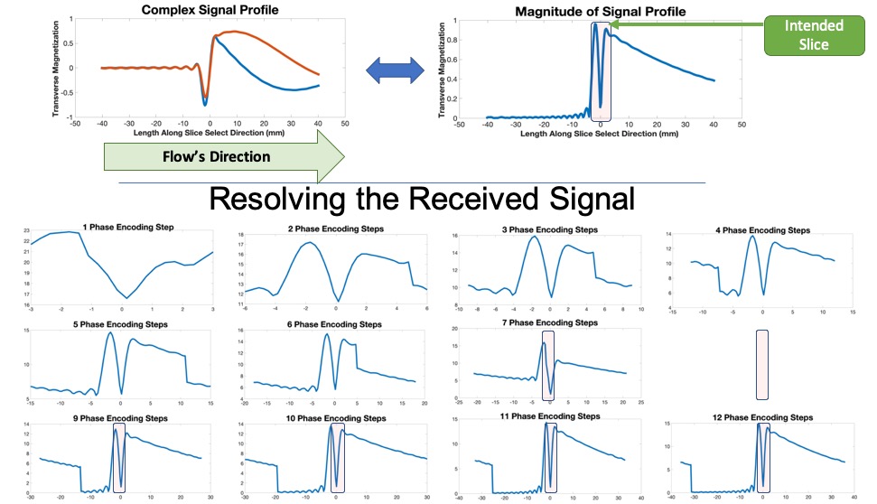
As shown in Fig.1 , when targeting a particular FOV that intersects a vessel the lack of spoiling preserves the outflowing signal that will only taper off via T*2 decay. The resulting equilibrium signal profile is sensitive to inflow-per-TR rate and off-resonance accumulation per TR ($$$\phi$$$), having considerable out-of-slice contributions from moving isochromats near $$$\pi$$$ rad/TR. Because bSSFP is prone to off-resonant-induced bands, the TR is often kept to less than 4ms. This keeps the decaying outflowing transverse magnetization persistent in following excitations. Because receivers cannot discriminate against this outflowing signal, it is projected onto the slice, resulting in the common outflow [16]. This provides us with a signal profile that expands beyond the nominal slice-thickness, spanning a 3D space. This is ultimately a special scenario of the aliasing phenomenon.Pulse Sequence
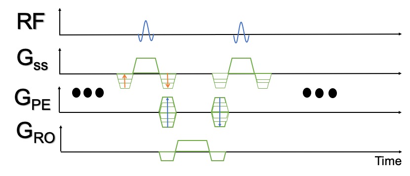
Fig. 2 Illustrates our proposed sequence, which includes the addition of balanced slice-encoding to resolve the outflowing signal. The encoding scheme was devised such that the resulting partitions would be just as thick as the anticipated slice. Therefore, with the same excitation profile, our proposed technique has an effective FOV that is a factor of the number of encoded partitions thicker than the standard 2D procedure. The slice-encoding resolves the space above and below the targeted slice, dedicating the center-partition to the intended FOV. Hence some aliasing is tolerated as long as it does not corrupt the center-partition, as demonstrated in Fig. 3 for a Bloch-simulated steady-state flowing signal profile.Methods
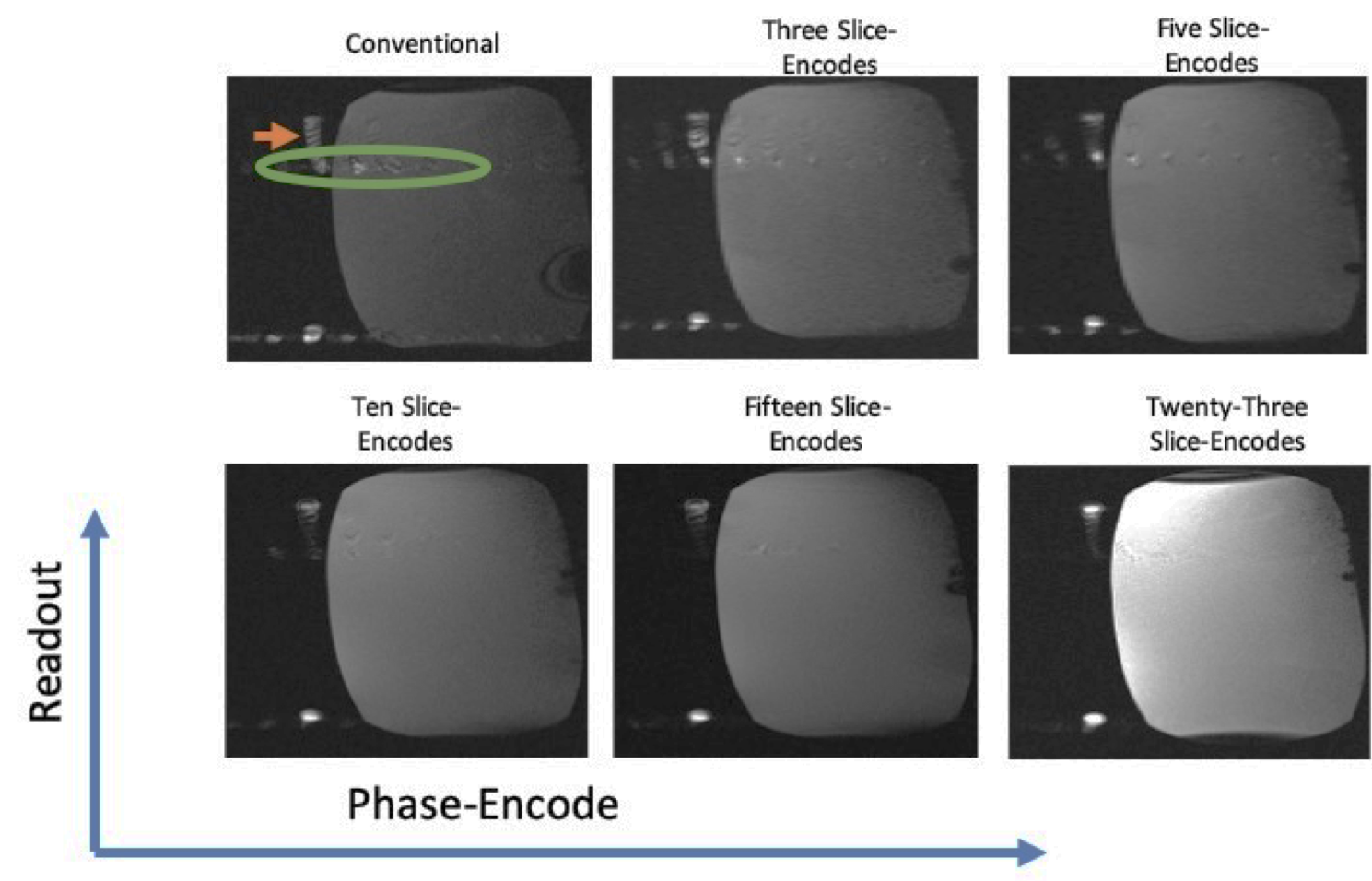
Simulation: A Bloch simulation to model steady-state signal with through-plane flow was implemented to investigate our method’s feasibility. Flow-velocity was modeled as the fraction of displaced spins per TR within an imaged slice (Ds). There was an array of spatially arranged spin-ensembles along the slice-select’s direction for settings for $$$\delta$$$s $$$\epsilon$$$ [0, 1] and $$$\delta$$$ $$$\phi$$$ $$$\epsilon$$$ [-2$$\pi\$$$p, 2$$$\pi$$$] The simulation was done for 300 excitations with the following parameters: T1/T2 values of 1000/150 ms, TE/TR = 2/4, 800ms sinc-functioned excitation-pulse, FA = 60°, TBW = 3, a $\pi$ rad/TR RF phase-ramp.Phantom Experiment: All investigations were performed on a 3T system, comparing standard 2D bSSFP scans with three, five, ten, fifteen, and twenty slice-encoded experiments. A homebrew flow phantom pumping tap-water with an average velocity of about 36 cm/s was used. Parameters were: TE/TR = 1.58/3.61 ms, 977 Hz/pixel readout-bandwidth, FOV = 330 x 196 mm, 146 x 256 in-plane matrix, and FA = 60°.
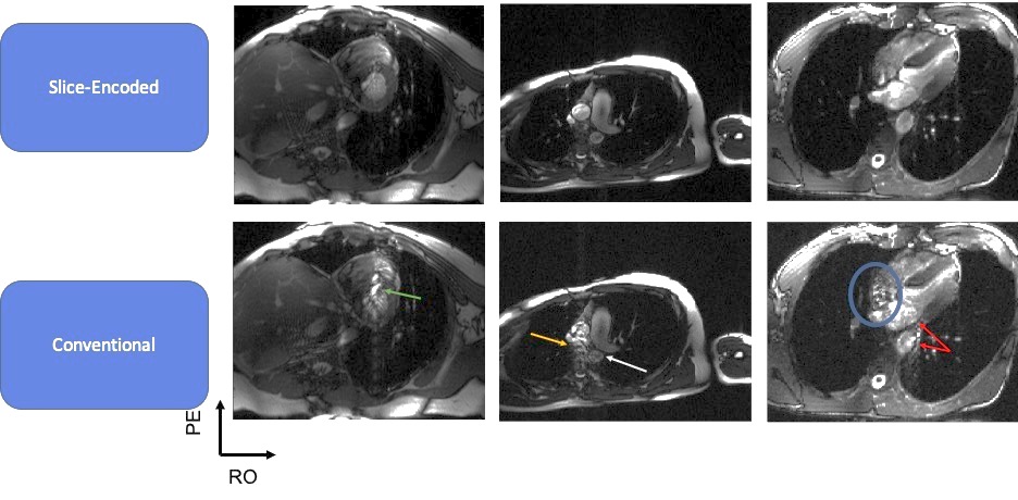
In Vivo Studies: Standard 2D and six slice-encoded cardiac-gated cine acquisitions were acquired for horizontal long axis (HLA) and short axis (SA) views on a 3T system from healthy volunteers ranging from ages 23-35. Parameters were: TE/TR = 1.58/3.61 ms, 977 Hz/pixel readout bandwidth, FA = 50°, a 1.4x1.4 mm in-plane resolution, a 256x184 in-plane matrix, GRAPPA-2X, and 6/8 partial Fourier, and 16 views-per-segment. Scans were achieved within a 20s breath-hold for the slice-encoded acquisition and 5s for the standard routine.
Results
Fig. 4 displays the flow-phantom experiments which demonstrate that outflow-sourced artifacts in the conventional acquisition are reduced with increasing number of slice-encoding steps. In vivo results are shown in Fig. 5, which compare three pairs of standard 2D with six slice-encoded acquisitions at peak systole. Each of the conventional scans are corrupted by signal pileups, signal misregistration, and a ghosting-like artifact that diminishes the myocardial-wall definition, vessel-borders, and clear chamber-visualization. These effects were significantly reduced upon adding slice-encoding steps.Discussion and Conclusion
We implemented a slice-encoding scheme to localize outflowing signal in an attempt to reduce flow-related artifacts commonly seen in cardiac cine bSSFP imaging. Significant improvements are made in the in vivo scans with even six partitions. The fact that this was achieved within a twenty-second breath-hold shows that this is possible to be incorporated within clinical routine in the foreseeable future, especially if it is integrated with a commonly undersampling strategy. This is particularly useful 3T and higher field strengths, given that these systems are prone to off-resonant effects.Acknowledgements
No acknowledgement found.References
1. Hawkes RC, Holland GN, Moor WS, Roebuck EJ, Worthington BS. Nuclear Magnetic Resonance (NMR) Tomography of the Normal Heart. J Comput Assist Tomogr 1981; 5(5): 605-612
2. Atkinson DJ, Edelman RRs. Cineangiography of the Heart in a Single Breath Hold with Segmented turboFLASH Sequence. Radiology 1991; 178(2):357-360
3. Patz S. Steady-State Free Precession: An Overview of Basic Concepts and Applications. Adv Magn Reson Imaging 1989; 1:73-102.
4. Wehrle FW. Fast-Scan Magnetic Resonanz: Principles and Applications. Magn. Reson Q 1990; 6: 165-236.
5. Haacke EM, Frahm J. Editorial; A Guide to Understanding Key Aspects of Fast Gradient Echo Imaging. JMRI 1992; 1:621-624.
6. Haack EM. Tach JA. Fast MR Imaging: Techniques and Clinical Applications. AJR 1990; 155:951-964.
7. Haacke EM, Wielopolski PA, Tkach JA. A Comprehensive Technical Review of Short TR Fast Magnetic Resonance Imaging. Rev Magn Reson Med 1991; 3:53-169
8. Scheffler, K. (1999), A Pictorial Description of Steady-States in Rapid Magnetic Resonance Imaging. Concepts Magn. Reson., 11:291-304. Doing: 10.1002/(SICI) 1099-0534(1999)11:5<291::AID-CMR2>3.0.CO;2-J
9. Scheffler, K. and Hennig, J. (2003), Is TrueFISP a Gradient-Echo or a Spin-Echo Sequence?. Magn. Reson. Med., 49: 395-397. Doi: 10.1002/mrm.10351
10. Maxcarenhas NB, Muthupillai R, Cheong B, Pereyra M, Flam,m SD. Fast 3D cine Steady-State Free Precession Imaging with Sensitivity Encoding for Assessment of Left Ventricular Function in a Single Breath-Hold. AJR Am J Roentgenol 2006; 187:1235-1239.
11. Nehrke K, Bornert P, Stehning C. Flow-Related Problems in Whole Heart Cine Coronary MR Angiography. Proceedings of 14th Scientific Annual Meeting of the ISMRM, Seattle, 2006. (abstract 2153).
12. Pfeffer MA, Braunwald E, Mode LA, et al. Effect of Captopril on Mortality and Morbidity in Patients with Left Ventricular Dysfunction After Myocardial Infarction: Results of the Survival and Ventricular Enlargement Trial. N Engl J Med 1992; 327:669-677.
13. Bonow RO, Lakatos E, Maron BJ, Epstein SE. Serial Long-Term Assessment of the Natural History of Asymptomatic Patients with Chronic Aortic Regurgitation and Normal Left Ventricular Systolic Function. Circulation 1991; 84: 1625-1635.
14. Verdecchia P, Schillaci G, Borgioni C, et Al Prognostic Significance of Serial Changes in Left Ventricular Mass in Essential Hypertension. Circulation 1998; 97:48-54.
15. Markl, M. , Alley, M. , Elkins, C. and Pelc, N. (2003), Flow Effects in Balanced Steady State Free Precession Imaging, Magn Reson. Med., 50: 892-903, dos: 10.1002/mrm.10631
16. Markl, M. and Pelc, N. J. (2004), On Flow Effects in Balanced Steady-State Free Precession Imaging: Pictorial Description, Parameter Dependence, and Clinical Implications. J. Mine. Reson. Imaging, 20: 697-705. Doing: 10.1002/jmri.20163
17. Storey P, Li W, Chen Q, Edelman RR. Flow Artifacts in Steady-State Free Precession Cine Imaging. Magn. Reson Med 2004; 51: 115-122.
18. Lu, W. Pauly, K. B., G. E., Pauly, J., M. and Hargreaves, B. A. (2009), SEMAC: Slice Encoding for Metal Artifact Correction in MRI. Magn. Reson. Med., 62: 66-76. doi: 10.1002/mrm.21967
Figures
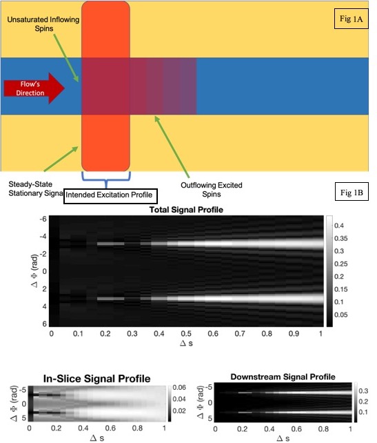
A-A 2D bSSFP acquisition in the presence of through-plane flow. Flowing spins only receive excitations while in the slice’s FOV. Their signal persists in bSSFP because it they’re not spoiled. B-The equilibrium signal-profile for $$$\Delta\Phi$$$ [-2$$$\pi$$$, 2$$$\pi$$$] and $$$\Delta\$$$s [0, 1]. The results include all spins simulated (top), spins within the slice’s FOV (bottom left), and all outflowed spins (bottom right). A clear off-resonant dependent phase accumulation exists, with the diagrams indicating enhancement is from outflowed spins.