2240
RF Coil and Gradient Shim Considerations for Hyperpolarized 13C Imaging of the Human Myocardium1GE Healthcare, Dallas, TX, United States, 2University of Texas Southwestern Medical Center, Dallas, TX, United States, 3GE Healthcare, Munich, Germany, 4GE Healthcare, Toronto, ON, Canada
Synopsis
Initial human hyperpolarized 13C cardiac imaging experiments utilized a frequency-selective, single-shot spiral pulse sequence for encoding, and a rigid 8 channel receiver array paired with a Helmholtz clamshell transmitter. In phantom studies, we evaluate this RF hardware along with flexible transmit / receiver designs and test these in healthy volunteers. Furthermore, as pointed out previously, the detected circumferential distribution of [13C]bicarbonate around the myocardium is often non-uniform in pigs, thus confounding the translation of this technique for regional metabolism evaluation. We observed this artifact in human volunteers and explore its possible sources.
Introduction
Understanding the role of altered intermediary metabolism in heart disease is important to addressing diverse clinical questions and new opportunities in therapy. In vivo assessment of cardiac metabolism in human subjects is critical. Earlier studies demonstrated imaging of [13C]bicarbonate in the human myocardium, a direct index of pyruvate oxidation in the citric acid cycle [1]. Initial imaging studies utilized an MRI pulse sequence consisting of a frequency-selective, single shot spiral in which one or more slices were encoded during mid-diastole [2]. This acquisition was paired with the clamshell-geometry radio frequency (RF) excitation Helmholtz coil [3] and the rigid, 8-channel paddle receivers. In this study, we evaluated two additional transmit / receive RF coils for 13C imaging over larger fields of view (FOV); initial transmit B1 profiles, SNR profiles, and in vivo images are provided. As pointed out in reference [4], the detected circumferential distribution of [13C]bicarbonate in the myocardium is often non-uniform in pigs, thus confounding the translation of this technique for regional metabolism evaluation. We observed this artifact in human volunteers and explore its possible sources.Methods
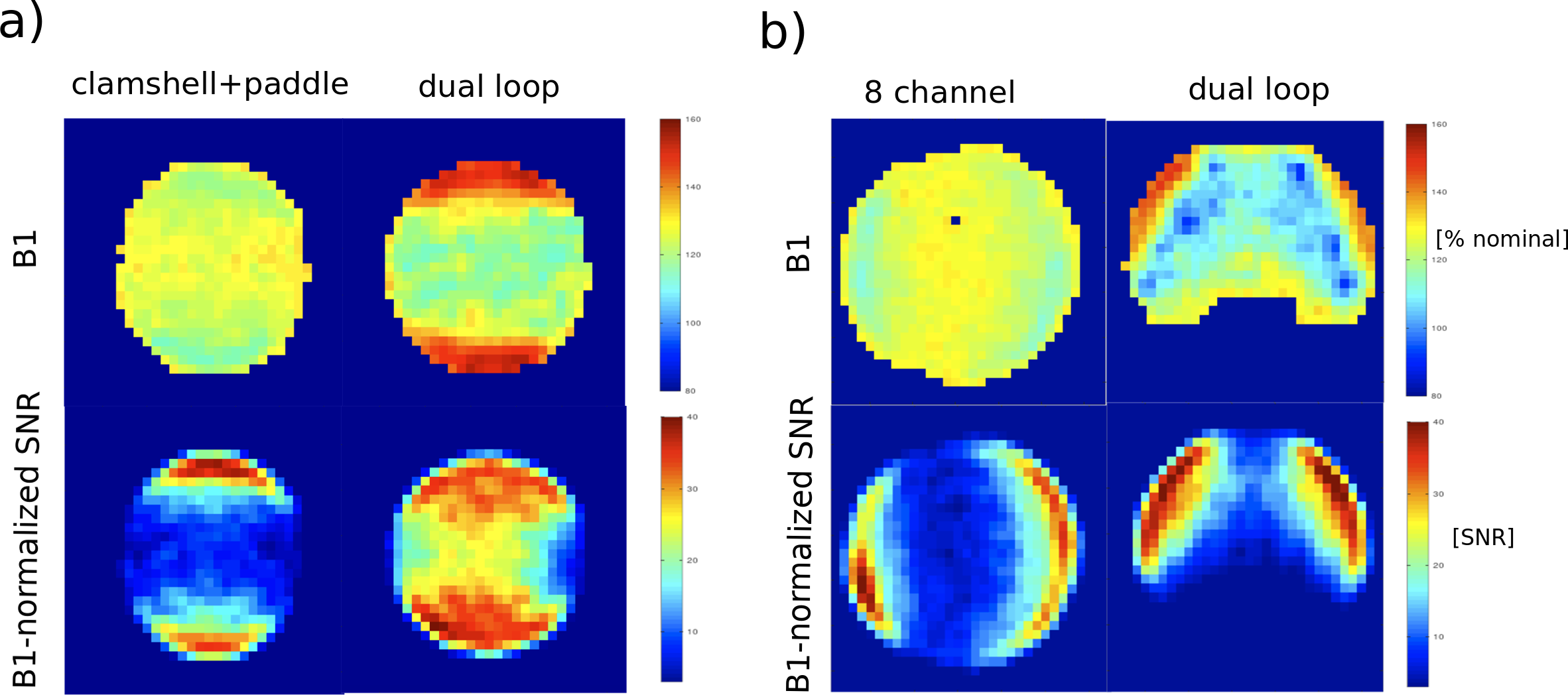
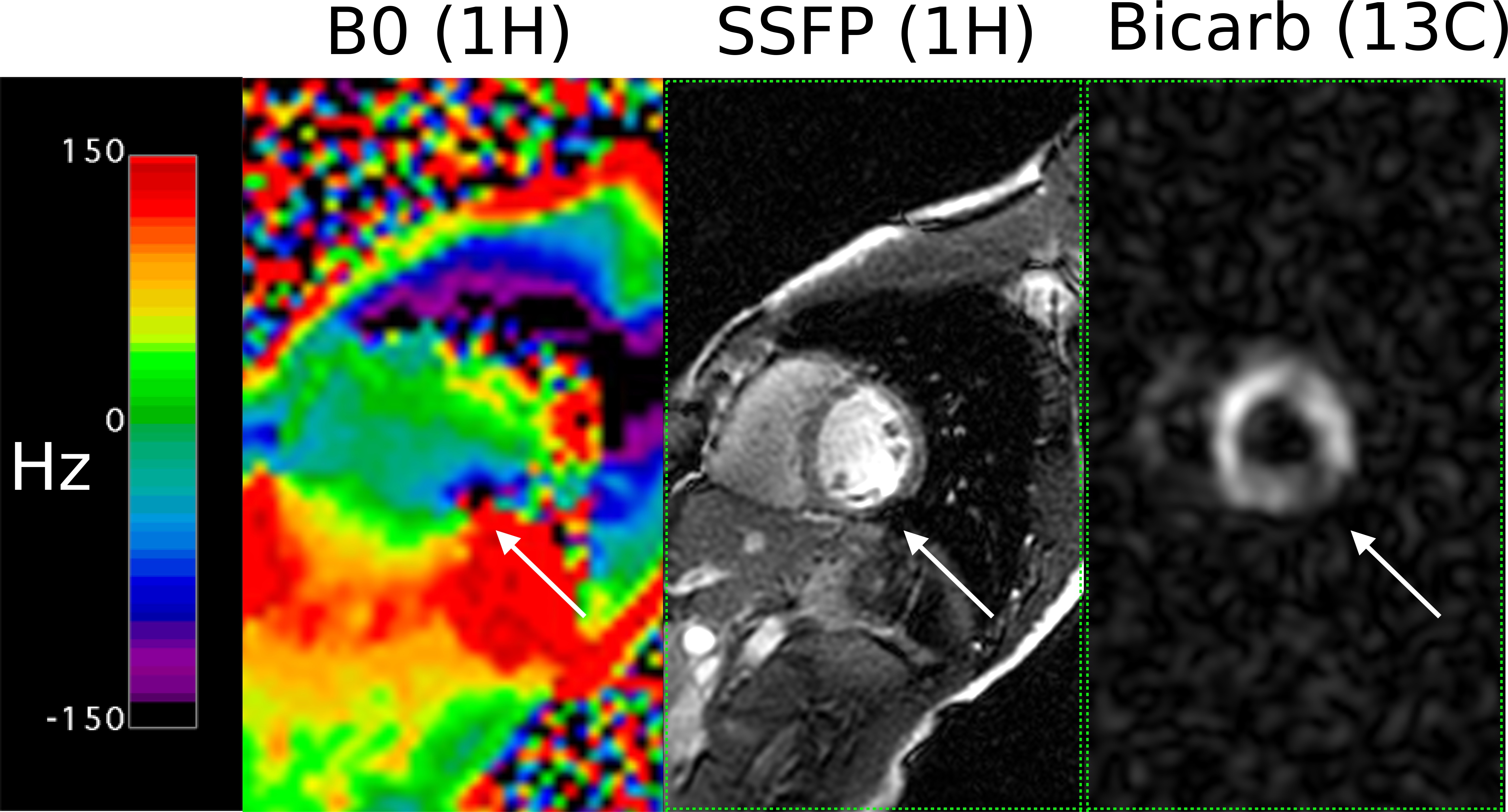
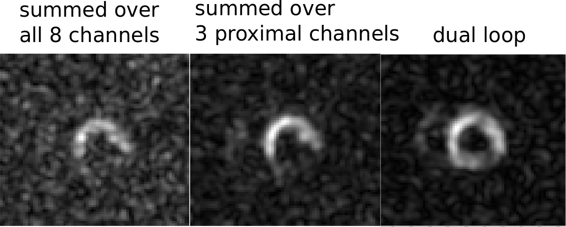
Three 13C-tuned RF transmit / receive configurations were tested to determine sensitivity, spatial coverage, and transmitter homogeneity. A clamshell transmitter / 8 channel rigid paddle [5] (GE Healthcare, Waukesha, WI), a dual 20 cm loop (Pulseteq, Chobham, UK) transceiver, and a flexible 8 channel array / vest transmitter (Clinical MR Solutions, Brookfield, WI) were measured using the SNR-measurement protocol, described in reference [6]. The protocol uses the natural abundance 13CH3 signal of the widely-distributed dimethyl silicone phantoms. A saline-filled ring enclosing the phantom provided RF loading. 2 sets of experiments were performed. In the first set, both the clamshell and dual loop coils were measured over a 32-cm FOV in a smaller 16 cm diameter phantom. In the second set of experiments, the flexible 8 channel and dual loop coils were measured over a 48-cm FOV in a larger 22 cm diameter phantom. Images were acquired at approximate flip angles of 45 and 90 degrees, 4s TR, and an flip angle map was reconstructed. The 90 degree image signal was then normalized by the sine of the estimated flip angle and noise to create a B1-normalized sensitivity map. Human Imaging and Dynamic Nuclear Polarization Subjects were scanned on a GE MR750w MRI system. The gradient shim values were calculated using a 14 cm FOV rectangular region enclosing the entire heart. A 1H imaging protocol including short-axis SSFP localizers and B0 maps were acquired with the body coil. A pre-determined scaling was applied to determine the center frequency shift for in vivo [1-13C]lactate based on in vivo 1H water resonant frequency. A cardiac-triggered, spectral-spatial excite (3 cm slice thickness), spiral readout gradient echo was adapted within the multi-nuclear research pack framework [7]. The 50 ms spiral readout had a nominal 40 cm FOV, 1.2 cm resolution, peak gradient amplitude 2.3 G/cm, and ramp rate of 74 G/cm/ms to conform to the restrictions of the wide bore gradient system [1-13C]pyruvate polarization and QC were performed via SPINLab polarizer. Injection was carried out over 12 seconds, and imaging initiated at the end of the infusion. One volunteer was imaged with both the dual loop and the flexible 8 channel array.Results
Figure 1 shows flip angle and SNR maps acquired in phantoms of the 3 coils studied. Due to its smaller transmitter size, the dual loop showed much higher transmitter inhomogeneity than either the clamshell or the 8-channel flexible array. Both the 8-channel array and dual loop have larger diameter receive elements, thus generating more uniform SNR profiles. The profile of these coils at a depth within the phantom typical of posterior left ventricle wall (15-20 cm) is at least 2x higher in both the dual loop and flexible 8-channel compared to the clamshell. Cardiac B0 maps, SSFP localizer, and [13C]bicarbonate are displayed in Figure 2. Note that the off-resonant region in the B0 map corresponds to an SSFP band as well as dropout in the [13C]bicarbonate image. [13C]bicarbonate images from the same volunteer using both the dual loop and the flexible 8 channel array are shown in Figure 3. SNR of the 8-channel array is comparable to the dual loop only when a small subset of channels proximal to the anterior chest wall are used.Discussion
Phantom measurements suggest that for imaging deeper tissue regions, the smaller receive elements of paddle receiver were suboptimal. The 8-channel array and the dual loop show comparable SNR distally, with the latter having a more inhomogeneous B1 field. The 8-channel showed comparable SNR in vivo to the dual loop only when a proximal subset of channels was selected, and this process can easily be automated via coil subspace-selection methods such as singular value decomposition [8]. Similar to the results in reference [4] in pigs, we observed non-uniform circumferential [13C]bicarbonate distribution in the myocardium when viewed on short axis. B0 mapping and SSFP imaging indicate that this is due to sup-optimal shimming, thus suggesting the need for improved B0 shim routines.Acknowledgements
No acknowledgement found.References
[1] C. H. Cunningham et al., “Hyperpolarized 13 C Metabolic MRI of the Human Heart,” Circ. Res., vol. 119, no. 11, pp. 1177–1182, Nov. 2016.
[2] A. Z. Lau et al., “Rapid multislice imaging of hyperpolarized 13C pyruvate and bicarbonate in the heart.,” Magn. Reson. Med., vol. 64, no. 5, pp. 1323–31, Nov. 2010.
[3] S. J. Nelson et al., “Metabolic Imaging of Patients with Prostate Cancer Using Hyperpolarized [1-13C]Pyruvate,” Sci. Transl. Med., vol. 5, no. 198, pp. 198ra108-198ra108, Aug. 2013.
[4] A. Z. Lau, A. P. Chen, and C. H. Cunningham, “Imaging the circumferential hyperpolarized 13C-bicarbonate distribution in the normal heart,” in International Society of Magnetic Resonance in Medicine, 2019, p. 0256.
[5] J. Tropp et al., “A carbon receive array of 8 elements, interoperable with proton scanning, for human temporal lobe,” in International Society of Magnetic Resonance in Medicine, 2012, p. 2658.
[6] G. D. Reed, R. F. Schulte, J. M. Park, C. R. Malloy, R. B. Hansen, and A. P. Chen, “Protocol for multi-site quantitative evaluation of 13C radio frequency coils,” in International Society of Magnetic Resonance in Medicine, 2019, p. 2510.
[7] F. Wiesinger et al., “IDEAL spiral CSI for dynamic metabolic MR imaging of hyperpolarized [1-13C]pyruvate,” Magn. Reson. Med., vol. 68, no. 1, pp. 8–16, Jul. 2012.
[8] Z. Zhu et al., “Coil combination methods for multi-channel hyperpolarized 13C imaging data from human studies,” J. Magn. Reson., vol. 301, pp. 73–79, Apr. 2019.
Figures