2221
Carotid Arterial Wall MRI Of ApoE-/- Mouse at 7T using 3D DANTE-Prepared Variable-Flip-Angle Rapid Acquisition with Relaxation Enhancement1Guangdong Provincial Key Laboratory of Medical Image Processing & Key Laboratory of Mental Health of the Ministry of Education, School of Biomedical Engineering, Southern Medical University, Guangzhou, China, 2Department of Pathophysiology, Key Lab for Shock and Microcirculation Research of Guangdong, School of Basic Medical Sciences, Southern Medical University, Guangzhou, 510515, China, Guangzhou, China, 3philips healthcare, Guangzhou, China, 4Department of Biomedical Engineering, School of Basic Sciences, Guangzhou Medical University, Guangzhou, China
Synopsis
Carotid atherosclerosis is a degenerative disease of the arterial wall, which can result in predisposition to cerebral thrombo-embolic stroke1. The combination of DANTE and VF-RARE (DANTE-VF-RARE) can suppress flow signal effectively. It was used for vessel wall imaging of human in the previous studies4. However, the feasibility of DANTE-VF-RARE sequence remains unclear for imaging of carotid for apoE-/- mouse. This work aims to develop 3D DANTE-VF-RARE on 7Tesla (T) and investigate its feasibility for vessel wall imaging of apoE-/- mouse.
Introduction
Carotid atherosclerosis is a degenerative disease of the arterial wall, which can result in predisposition to cerebral thrombo-embolic stroke1. Three-dimensional (3D) variable flip-angle Rapid Acquisition with Relaxation Enhancement (VF-RARE) sequence can achieve near constant echo amplitudes over long echo trains and has been demonstrated successfully in vessel wall imaging2,3. Meanwhile, the combination of DANTE and VF-RARE (DANTE-VF-RARE) can suppress flow signal effectively. It was used for vessel wall imaging of human in the previous studies4. However, the feasibility of DANTE-VF-RARE sequence remains unclear for imaging of carotid for apoE-/- mouse. This work aims to develop 3D DANTE-VF-RARE on 7Tesla (T) and investigate its feasibility for vessel wall imaging of apoE-/- mouse.Methods
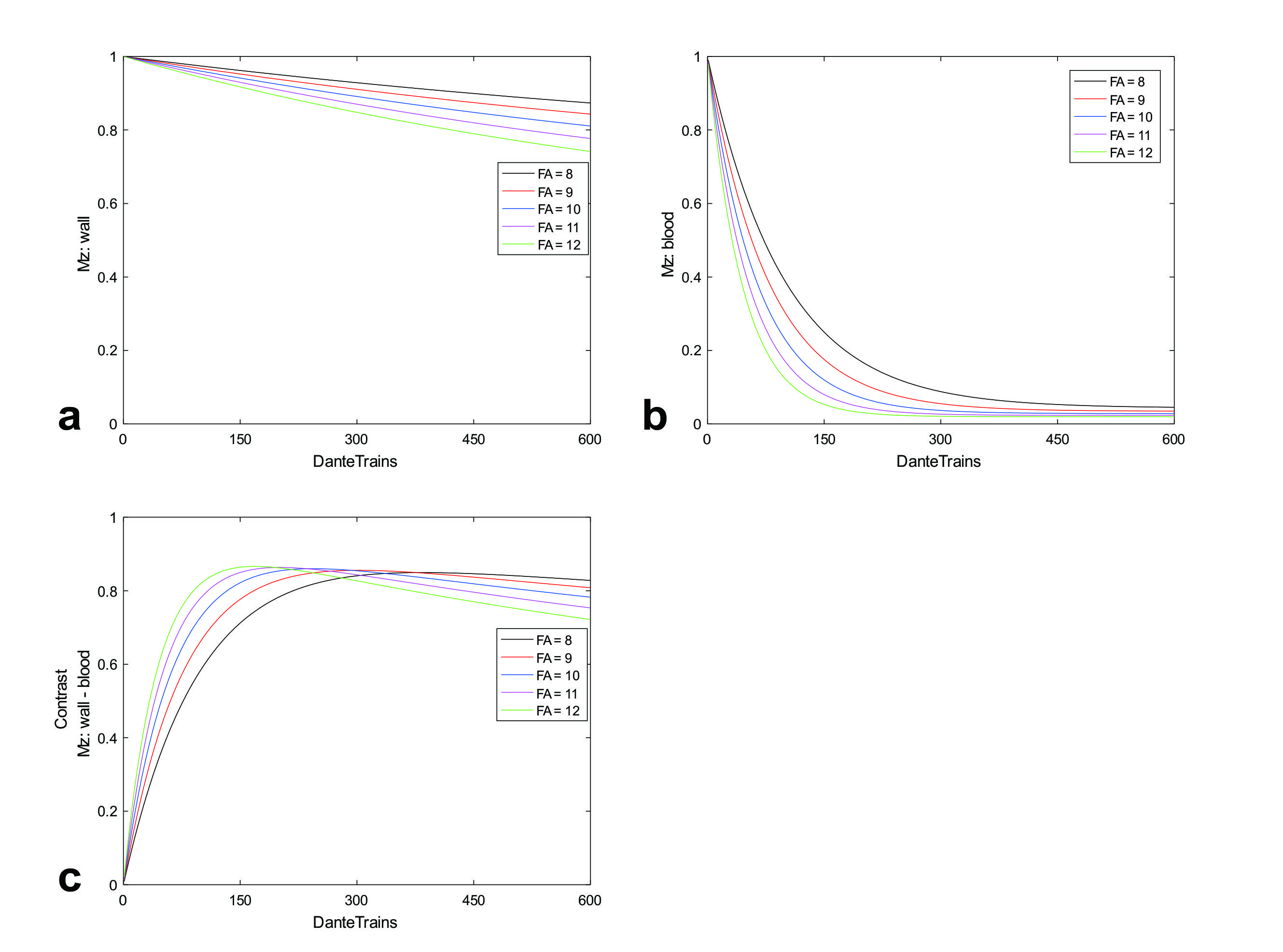
DANTE OptimizationThe simulation program was implemented in Matlab. The goal of the DANTE optimization is to maximize the contrast between the vessel wall and blood. In numerical simulations, the T1/T2 values of the vessel wall and arterial blood were 1628/46 ms and 2200/68 ms, respectively. The flip angle was ranged from with increment of 1o and the number of pulse was ranged from 0 to 600. To simplify the numerical simulations, an approximation signal evolution5 was adopted, so that the blood flow velocity was not considered.
Experiments
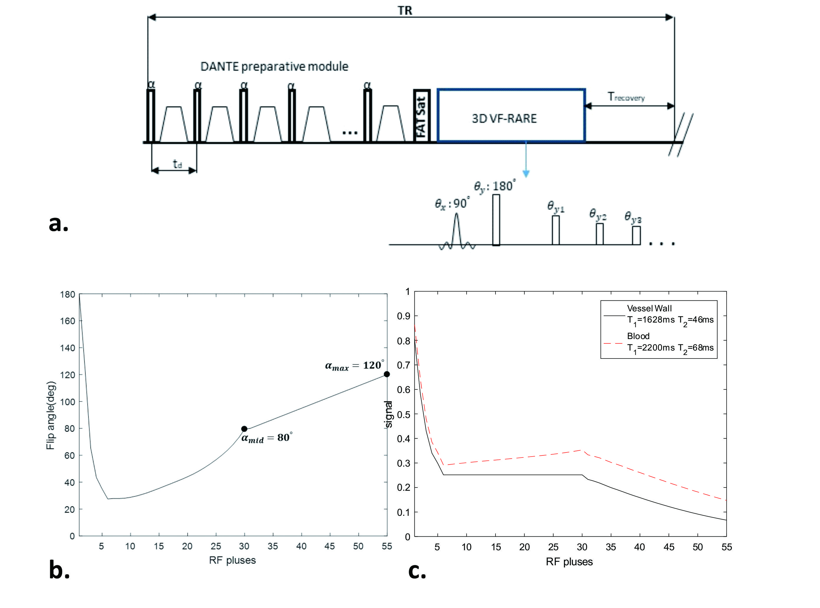
This study was approved by local institutional review board. Three ApoE-/- mice (8 weeks old) were fed with a high-fat diet for approximately 32 weeks. Mice underwent MR scan using Bruker 7T MRI scanner. Figure 1(a) shows a schematic representation of the DANTE-VF-RARE pulse sequence. The parameters for the DANTE module included: flip angle :10°, pulse train length :150, inter-pulse duration td: 0.97 ms. The parameters for the VF-RARE readout included: TR/TE = 1200/22.3 ms, FOV = 20×20×16 mm3, True resolution= 0.1×0.1×0.2 mm3 and reconstructed to 0.05×0.05×0.1 mm3, bandwidth=500 Hz/pixel, slice partial Fourier factor = 6/8, NA= 3, total scan time= 14 min24s. The variable-flip angle scheme for the VF-RARE acquisition is illustrated in Fig. 1(b). Conventional 2D RARE images were acquired as a reference with the following parameters: TR/TE = 1200/7 ms , FOV = 20×20 mm2 ,in-plane True resolution= 0.1×0.1 mm2 and reconstructed to 0.05×0.05 mm2; slice thickness = 0.5 mm, bandwidth=500 Hz/pixel; NA= 3, total scan time= 12 min. A pair of parallel saturation bands were placed above and below the slices group. To confirm structural changes of the carotid lumen detected by VF-RARE images, A FLASH sequence was performed for bright blood imaging with following imaging parameters : TR/TE = 15/2.6ms, flip angle = 20◦, FOV =20×20×16 mm3, True resolution:= 0.1×0.1×0.2 mm3 and reconstructed to 0.05×0.05×0.1 mm3, NA= 3, total scan time= 13min12s.
Histology
Two days after MRI measurements, histologic sections were made. Carotid artery was sectioned into 5-µm serial sections perpendicular to the vessel direction.
Image Analysis
ImageJ was used to measure lumen signal ( Slumen ), noise level( σ ) and wall signal(Swall). Swall was measured as the mean signal intensity of a one-pixel-width circumferential path traced manually through the middle of the arterial wall6. To allow for fair comparison of CNR values with respect to imaging parameter differences, CNR efficiency ( CNReff ) was calculated by the relation7:
CNReff=CNR÷(VOXEL×√TASLICE )
Where VOXEL is the imaging voxel volume and was expressed in cubic millimeters, TASLICE is the scan time for each slice (expressed in minutes).
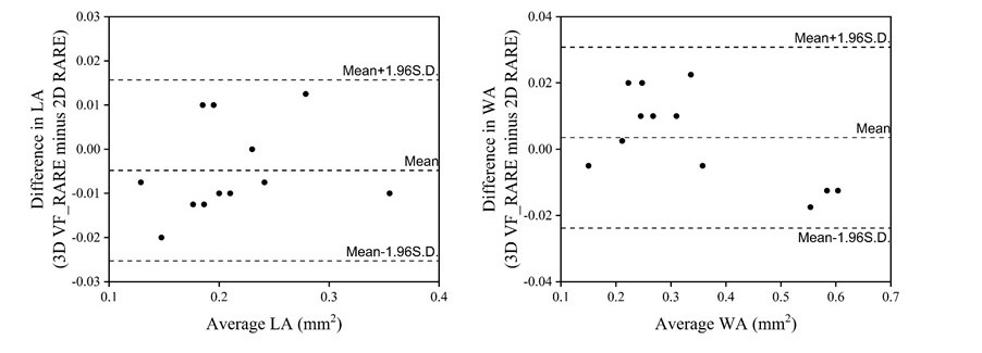
SPSS was used for statistical analysis. A two-tailed paired Student’s t test was performed between 3D DANTE-VF-RARE and 2D RARE images for SNRlumen, SNRlumen , CNRwall_lumen , CNReff , LA(carotid lumen area), WA(carotid wall area); Moreover, the intra-class correlation coefficient (ICC) and Bland-Altman plots were performed to assess the agreement in WA and LA measurements obtained from the 3D DANTE-VF-RARE and 2D RARE . A p value of less than 0.05 was considered statistically significant. Data were presented as means standard deviations.
Result
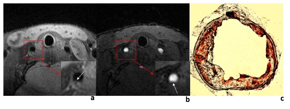
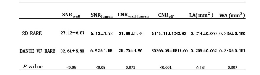
Figure 3c shows that the contrast is anticipate to peak earlier but in a lower steady state for higher flip angles, as compared with that for lower flip angles. Figs. 3 shows that sufficient contrast can be obtained using a DANTE pulse with flip angle of 10° and the train length of approximately 150. 3D DANTE-VF-RARE and 2D RARE images from a representative ApoE-/- mouse are shown in Figs. 3 and 4. DANTE-VF-RARE can generate a comparable image quality while achieving 2.5 fold improvement in slice resolution over 2D RARE (0.2mm vs 0.5mm). Quantitative measurements and statistical analyses were listed in Table 1. There were no significant difference in CNRwall_lumen (25.70±4.96 vs 21.99±5.34, P=0.071) between the 3D and 2D acquisitions. However, the 3D protocol offered a significantly improved CNReff (30266.98 ± 5844.60 vs 5115.11±1242.83, P<0.001). For a comparison of morphology, the carotid LA and WA obtained from DANTE-VF-RARE and 2D RARE were in a good agreement based on the ICC analysis (LA: ICC=0.983; WA: ICC =0.996) and the Bland-Altman plots(Figs.5).Discussion and Conclusion
In this work, the results shows that the combination of DANTE and VF-RARE can achieve high spatial resolution carotid artery imaging at 7T. 3D DANTE-VF-RARE is able to generate a comparable while achieving 2.5 fold improvement in slice resolution over 2D RARE (0.2mm vs 0.5mm). 3D DANTE-VF_RARE allows for reliable visualization and quantitative evaluation of the arteriosclerotic lesions within the carotid artery of an apoE-/- mouse model at 7T.Acknowledgements
No acknowledgement found.References
1.Ingall T . Stroke-incidence, mortality, morbidity and risk[J]. Journal of insurance medicine (New York, N.Y.),2004, 36(2):143-152.
2. Fan Z , Zhang Z , Chung Y C , et al. Carotid arterial wall MRI at 3T using 3D variable-flip-angle turbo spin-echo (TSE) with flow-sensitive dephasing (FSD)[J]. Journal of Magnetic Resonance Imaging Jmri, 2010, 31(3):645-654.
3.Qiao Y , Steinman D A , Qin Q , et al. Intracranial arterial wall imaging using three-dimensional high isotropic resolution black blood MRI at 3.0 Tesla[J]. Journal of magnetic resonance imaging : JMRI, 2011, 34(1):22-30.
4.Xie Y , Yang Q , Xie G , et al. Improved black-blood imaging using DANTE-SPACE for combined carotid and intracranial vessel wall evaluation[J]. Magnetic Resonance in Medicine, 2015, 17(Suppl 1):2286-2294.
5.Li L, Miller KL, Jezzard P. DANTE-prepared pulse trains: a novel approach to motion-sensitized and motion-suppressed quantitative magnetic resonance imaging. Magn Reson Med 2012;68: 1423–1438
6.Koktzoglou I , Li D . Diffusion-Prepared Segmented Steady-State Free Precession: Application to 3D Black-Blood Cardiovascular Magnetic Resonance of the Thoracic Aorta and Carotid Artery Walls[J]. Journal of Cardiovascular Magnetic Resonance, 2007, 9(1):33-42.
7.Zhang Z, Fan Z, Carroll TJ, et al. Three-dimensional T2-weighted MRI of the human femoral arterial vessel wall at 3.0 Tesla. Invest Radiol 2009;44:619–626
Figures