2201
Clinical value of Cardiac Magnetic Resonance Imaging in Primary Cardiac tumors1Department of Radiology, Beijing Anzhen Hospital, Capital Medical University, Beijing, China, 2Philips Healthcare, Beijing, China
Synopsis
Cardiac magnetic resonance (CMR) offers superior advantages in cardiac imaging because of greater field of view, excellent soft-tissue imaging, and multiplanar imaging capabilities. CMR imaging can evaluate the characteristics of cardiac tumors by demonstrating the relationship between the tumor and its surrounding tissues,. Moreover, it plays a significant role in assisting the formulation of the surgical plan, in addition to the assessment of tumor progression and the monitoring of postoperative tumor recurrence and metastasis. Our objective is to determine the value of CMR in assessing the likelihood of primary cardiac tumors and guiding patient management.
Synopsis
Cardiac magnetic resonance (CMR) offers superior advantages in cardiac imaging because of greater field of view, excellent soft-tissue imaging, and multiplanar imaging capabilities. CMR imaging can evaluate the characteristics of cardiac tumors by demonstrating the relationship between the tumor and its surrounding tissues,. Moreover, it plays a significant role in assisting the formulation of the surgical plan, in addition to the assessment of tumor progression and the monitoring of postoperative tumor recurrence and metastasis. Our objective is to determine the value of CMR in assessing the likelihood of primary cardiac tumors and guiding patient management.Introduction
Cardiac masses include tumors and non-neoplastic lesions[1-2]. Cardiac tumors can be characterized by masses in the cardiac cavity, myocardium or pericardium, which can involve valves or papillary muscles[3]. In 2015, the World Health Organization (WHO) released a new histologic classification of primary cardiac tumors which are divided into benign tumors, uncertain biological behavior tumors, germ cell tumors and malignant tumors[4-5]. Echocardiography plays a key role in the diagnosis of heart tumors, but echocardiography can not accurately analyze the characteristics of tumors and distinguish between benign and malignant tumors. Cardiac magnetic resonance imaging (CMR) is a multi-parameter imaging technique, which is currently considered to be the gold standard for non-invasive display of soft tissue characteristics. In addition, CMR can facilitate the choice of surgical procedures by providing information about cardiac structure and function as well as hemodynamic impairment[6]. According to CMR pocket guide, the standard protocols include: high resolution anatomy, cine imaging in all standard and targeted planes, black-blood T1W, T2W, first pass perfusion and LGE. In this study, we analyzed the CMR features, clinical and pathological characteristics of the most common primary cardiac tumors in order to discuss the significance of CMR imaging in the diagnosis, treatment and prognosis of cardiac tumors[7-8].Material and Methods
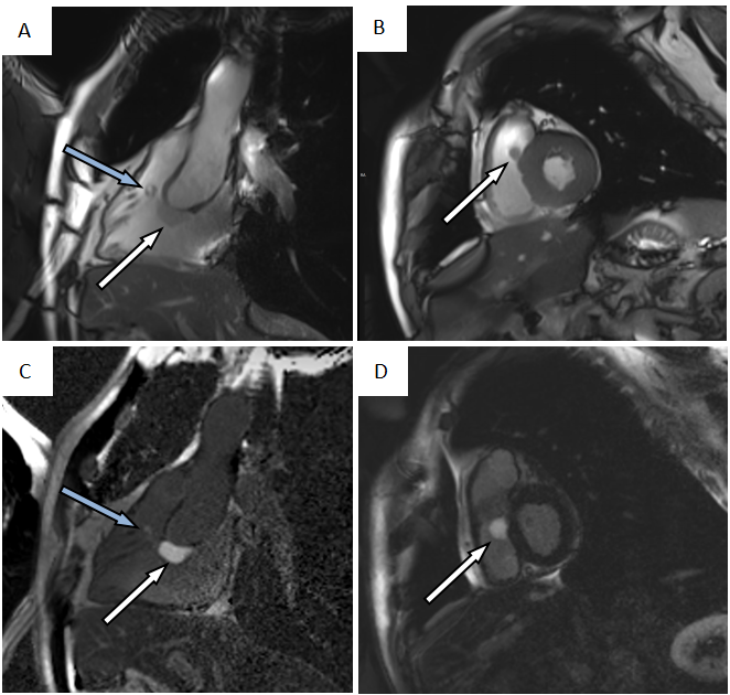
We retrospectively analyzed cardiac tumors patients who underwent CMR from January 2007 to June 2019 in scanners from multiple vendors. The patients were evaluated comprehensively from the aspects of sex, age, imaging manifestations, complications, pathological results and laboratory indexes. The classification, clinical manifestations, CMR features, treatment and prognosis of cardiac tumors were described. The MR characteristics of cardiac tumor patients in cine images, T1-weighted imaging (T1W) and T2-weighted imaging (T2W), contrast first-pass perfusion (FPP), post-contrast inversion time (TI) scout and late gadolinium enhanced (LGE) sequence were analyzed. 169 patients with cardiac tumors were included for analysis. Tumors’ localization, tissue signal, primary tumor characteristics, secondary changes and extracardiac findings were studied.Results
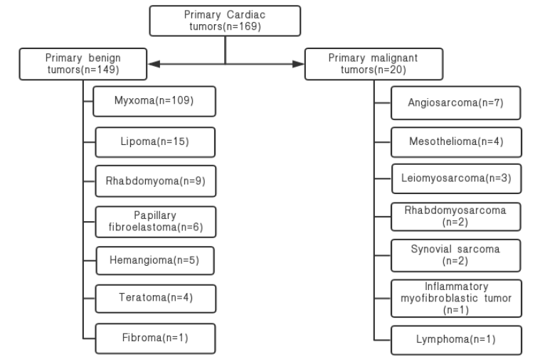
We collected about 169 patients with cardiac tumors in a single center in the past twelve years shows in figure 1, including 63 males and 106 females, with a male to female ratio of 0.59. The age ranges from 1 to 75 years old, with an average age of 38 years. According to the WHO Classification of Cardiac tumors, there were 149 cases of benign tumors(88%), 20 cases of malignant tumors(12%). Among benign tumors, myxoma was the most common. Angiosarcoma is the most common malignant tumor about 7 cases (4.1%). The location of benign tumors was frequently seen in the left atrium, about 106 cases (71.1%), malignant tumors were more common in the right heart system, and the right atrium accounting for about 15 cases (75%). Benign tumors are usually solitary, well-defined, regular in shape, small in size and non-invasive to surrounding tissues. Malignant tumors are more prone to larger than 5cm or multiple, with ill-defined border and irregular shape. The tumor directly infiltrates the pericardium and its surrounding structure, which can lead to a large number of pericardial effusion and pericardial nodules.Discussion
This study shows the unique CMR features of cardiac tumors which is contribute to the accurate evaluation before operation, so as to avoid unnecessary cardiac biopsies. Many cardiac masses are found by accident in routine echocardiography. However, there are some limitations in echocardiography, including the dependence of the operator, limited visual field (especially in patients with lung disease or larger body habitus), and restricted imaging of the right heart, mediastinum and extracardiac structures. MR imaging provides higher time resolution and better tissue characterization, multi-directional imaging and does not expose patients to ionizing radiation. CMR imaging can reliably detect thrombus and has been proved to be able to accurately distinguish cardiac non-tumor lesions, benign tumors and malignant tumors. CMR can distinguish between different tissue characteristics, such as water and fat content, which leads to specific signal patterns on T1WI and T2WI. Contrast enhancement can be used to demonstrate additional tissue properties, such as vascularity and fibrosis. CMR can be used to monitor tumor regression after surgery or radiotherapy and chemotherapy. The morphological, anatomical, histological characteristics and functional effects of suspected tumors can be evaluated in a single CMR examination. CMR imaging is becoming an established method for further assessment in cardiac masses.Conclusion
The fundamental role of CMR in the treatment of cardiac masses depends on accurate localization and qualitative diagnosis. CMR can provide accurate differentiation of pseudomasses, benign and malignant masses. It is an ideal tool for preoperative evaluation and follow-up in patients with cardiac tumors, proving superior to echocardiography in establishing the type of cardiac tumour and planning management.Acknowledgements
No acknowledgement found.References
1. Wang JG. Primary Cardiac Tumors. Right Heart Pathol 2018;489-514.
2. Carrascosa PM, Rodríguez-Granillo GA, Deviggiano A, et al. Cardiac Masses and Tumors. Clinical Atlas of Cardiac and Aortic CT and MRI 2019;287-307.
3. Butany J, Nair V, Naseemuddin A, et al. Cardiac tumours:diagnosis and management. Lancet Oncol 2005;6:219-28.
4. Jain S, J Maleszewski J, Christopher R, et al. Current diagnosis and management of cardiac myxomas. Expert Rev Cardiovasc Ther 2015;13:369-75.
5. Poterucha TJ, Kochav J, O’Connor DS, et al. Cardiac Tumors: Clinical Presentation, Diagnosis, and Management. Curr Treat Options in Oncol 2019;20:66.
6. Beroukhim RS, Prakash A, Buechel ER, et al. Characterization of cardiac tumors in children by cardiovascular magnetic resonance imaging: a multicenter experience. J Am Coll Cardiol 2011;58:1044-54.
7. Sparrow P, Kurian J, Jones T, et al. MR imaging of cardiac tumors. Radiographics 2005;25:1255-76.
8. Motwani M, Kidambi A, Herzog BA, et al. MR imaging of Cardiac tumors and masses: A Review of Methods and Clinical Applications. Radiology 2013;268:26-43.
Figures