2195
Relationships between CMR, rheometry and Raman spectroscopy of doxorubicin induced cardiotoxicity in the swine.
Delphine Perie1, Eric Buzaglo1, Clemence Balosetti1, Helene Heon2, and Daniel Curnier3
1Mechanical Engineering, Polytechnique Montreal, Montreal, QC, Canada, 2Research Center, CHUM, Montreal, QC, Canada, 3Kinesiology, University of Montreal, Montreal, QC, Canada
1Mechanical Engineering, Polytechnique Montreal, Montreal, QC, Canada, 2Research Center, CHUM, Montreal, QC, Canada, 3Kinesiology, University of Montreal, Montreal, QC, Canada
Synopsis
CMR has already been used to investigate the long term effects of cancer treatments. We investigated relationships between clinical, CMR, mechanical and biochemical properties of the myocardium and their changes due to early doxorubicin induced cardiotoxicity in the swine model. MR relaxation times and CMR function parameters highlighted the importance of regional analysis because of the heterogeneity in the tissue response to doxorubicin treatment. The observation of different remodelling patterns between moving and fixed cardiac walls needs to be translated to personalized diagnosis and medicine approaches for cancer treatment application.
Introduction
Cancer chemotherapy is an effective treatment in both adults and children. However, the associated cardiotoxicity of doxorubicin, the most commonly used anticancer agent, is a well-known serious side effect leading to long-term morbidity. Cardiovascular magnetic resonance (CMR) has already been used to investigate the long term effects of cancer treatments [1-3]. We previously presented an animal study of the early effects of doxorubicin on the swine model and its detection with a multiparametric CMR protocol [4]. Mechanical properties can be determined from multiparametric CMR data associated to principal component analysis [5]. Raman spectrometry is a promising cancer detection technique for surgical guidance applications, which provide quantifiable tissue properties associated to the structural, metabolic, immunological and genetic biochemical composition [6]. The aim of this study was to investigate relationships between clinical, CMR, mechanical and biochemical properties of the myocardium and their changes due to early doxorubicin induced cardiotoxicity in the swine model.Methods
The samples and the data used in this study were extracted from a previous animal experiment approved by our IRB [4]. In this previous experiment, five young female miniature swines received five 75mg/m2 injections of doxorubicin every three weeks for a total cumulative dose of 375mg/m2, constituting the Doxo group. Two miniature swines received a saline injection under the same protocol, constituting the Control group. All animals underwent their last CMR acquisition 2 weeks after the end of the treatment on a Siemens Skyra 3T MR system using a 18-channel phased array body matrix coil. The CMR protocol included a MOLLI sequence for T1 mapping, a T2-prepared TrueFISP sequence for T2 mapping at apical, mid-ventricular and basal levels (pixel resolution 1.4mmx1.4mmx8.0mm), and an ECG-gated cine TruFISP sequence (14 slices in short axis and 5 slices in long axis, slice thickness 8mm, repetition time 34.6ms, effective echo time 1.2ms, flip angle 38°, iPAT factor 3, matrix 208x210 and in-plane pixel size 1.25x1.25 mm). After this last CMR acquisition, the animals were sacrificed with a barbiturate overdose (108mg/kg) and myocardial samples were collected and preserved in liquid nitrogen. These samples were subjected to frequency sweep and stress-relaxation tests using a rheometer (Figure 1) in order to determine the shear modulus, loss modulus, storage modulus (Figure 2), damping factor, complex viscosity and relaxation modulus (Figure 3). Samples were also placed in a Raman probe after being submerged in a saline solution to determine the molecular composition from the Raman spectrum (Figure 4). Spearman correlation test were performed between clinical, CMR, mechanical and chemical properties of the myocardium using SigmaStat (Systat Software, Inc.).Results
Miniature swines receiving chemotherapy yielded differences in both CMR, mechanical and clinical properties (Figure 5). An overall decrease in the storage and loss modulus was observed in the septum while an overall strengthening of these parameters was observed in the left and right ventricles. The stress-relaxation test showed a clear drop in the mechanical properties of all Doxo samples (Figure 3). The Raman spectrometry results showed an increase of the collagen content in the left and right ventricles while a decrease of the collagen content was observed in the septum. The Raman spectrometry results also showed an overall decrease of the lipid content in the Doxo samples. Significant correlations (p<0.001) were found between T1 and EDV (r=0.83), relaxation modulus (r=0.81), damping factor (r=-0.67) or neutrophils (r=0.79), between T1gd and platelets (r=0.81) or complex viscosity (r=0.71), between ESV and relaxation modulus (r=0.76), between EDV and loss modulus (r=-0,68), damping factor (r=-0,64), white blood cells (r=0,87) or neutrophils (r=0.93), between platelets and relaxation modulus (r=0.81), storage modulus (r=0.75) or complex viscosity (r=0.81).Discussion
Both imaging (CMR and Raman spectrometry) and mechanical (shear tests) data showed a decrease in the overall structural integrity associated to a lack of collagen in the cardiac fixed wall (septum) while increased structural integrity associated to fibrosis was found in the cardiac mobile wall (left ventricle) of the Doxo group. The myocardium remodeling during the doxorubicin treatment might be influenced by the mechanical stress withstood by the cardiac cells, which are different between the mobile and fixed walls of the ventricles. Both imaging and mechanical data showed a decreased shear relaxation modulus associated to a drop of the lipid content in both fixed and mobile walls of the Doxo group. These decreases might be associated to the weight loss and the progressive non-regenerative anemia developed by the animals of the Doxo group. Our previous study [4] showed that CMR parameters were able discriminate Doxo animals from controls and that these differences were detectible earlier than onset of classical echocardiographic changes. The proposed study confirms the relationship between the CMR parameters and the doxorubicin induced disturbed myocardium remodeling.Conclusion
MR relaxation times and CMR function parameters reflected both mechanical and biochemical changes within the myocardium in early doxorubicin induced cardiotoxicity. They also highlighted the importance of local or regional analysis because of the heterogeneity in the tissue response to doxorubicin treatment. The observation of different remodelling patterns between moving and fixed cardiac walls needs to be translated to personalized diagnosis and medicine approaches for cancer treatment application.Acknowledgements
NSERC, FRQNT and Polytechnique Montreal for the financial support, researchers from the people from the CRCHUM for their help on the animal care, data acquisition and data analysis.References
1- K. Ylanen, T. Poutanen, P. Savikurki-Heikkila, I. Rinta-Kiikka, A. Eerola, and K. Vettenranta, "Cardiac magnetic resonance imaging in the evaluation of the late effects of anthracyclines among long-term survivors of childhood cancer," J Am Coll Cardiol, vol. 61, pp. 1539-47, Apr 9 2013. 2- Armstrong GT, Plana JC, Zhang N, Srivastava D, Green DM, Ness KK, Daniel Donovan F, Metzger ML, Arevalo A, Durand JB, Joshi V, Hudson MM, Robison LL, Flamm SD. Screening adult survivors of childhood cancer for cardiomyopathy: comparison of echocardiography and cardiac magnetic resonance imaging. J Clin Oncol. 2012 : 30(23):2876-84. 3- Tham EB, Haykowsky MJ, Chow K, Spavor M, Kaneko S, Khoo NS, et al. Diffuse myocardial fibrosis by T1-mapping in children with subclinical anthracycline cardiotoxicity: relationship to exercise capacity, cumulative dose and remodeling. J Cardiovasc Magn Reson 2013, 15:48. 4- Périé D., Balosetti C., Héon H., Dahdah N., Cheriet F., Friedrich M.G., Curnier D. (2017), Multiparametric CMR protocol and analysis methods for the detection of early cardiotoxicity remodelling in the mini-swine. International Research Society on Magnetic Resonance in Medecine, Hawaï (USA). 5- Périé D, Dahdah N, Foudis A, Curnier D. Multiparametric MRI as an indirect evaluation tool of the mechanical properties of in-vitro cardiac tissues. BMC Cardiovasc Disord. 2013 27; 13:24. 6- I. Santos, E. Barroso, T. Bakker-Schut, “Raman spectroscopy for cancer detection and cancer surgery guidance: translation to the clinics,” Analyst, Issue 17, 2017.Figures
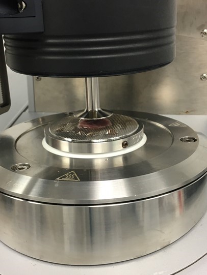
Figure 1: Rheometer
used for the shear frequency sweep and relaxation tests on the cardiac samples.
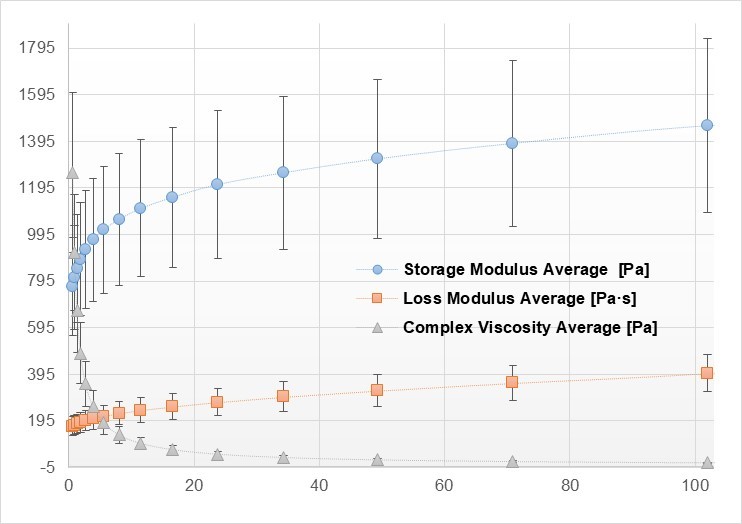
Figure
2: Storage modulus, loss modulus and complex viscosity for a frequency sweep test on a control
sample at 1.6% strain.
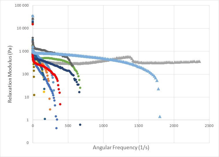
Figure
3: Relaxation Modulus for the doxo samples (shortest angular frequencies) and
control samples (longest angular frequencies).
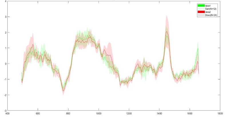
Figure
4: Raman spectrum obtained for the doxo sampels (red) and control samples
(green).
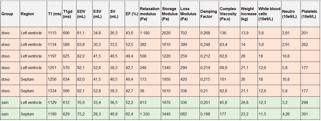
Figure 5: CMR (Relaxation times T1 and T1gd, function parameters EDV, ESV,
SV and EF), mechanical (relaxation modulus, storage modulus, loss modulus,
damping factor, complex viscosity) and clinical (weight increase, white blood
cells, neutrophils and platelets) results.