2189
Outer volume suppressed 3D-encoded DENSE MRI for quantifying biventricular myocardial strain during a breath-hold1Geisinger, Danville, PA, United States
Synopsis
Myocardial strain is a highly sensitive measure of heart function, but techniques to measure strain, such as displacement-encoded (DENSE) MRI, typically focus on the left ventricle, despite known clinical importance of right ventricle mechanics. The high resolution required to measure right ventricular strains is possible using navigator-based approaches; however, these are time-consuming and impractical for broad use. DENSE with outer volume suppression has been applied during a breath hold in the left ventricle, and may also enable resolving the right ventricle. We implemented DENSE with outer volume suppression and achieved similar 3D biventricular strain measurements compared to a navigator-based acquisition.
Background
Myocardial strain measurements are sensitive to changes in cardiac tissue structure and function during disease.1 The noninvasive measurement of strain is possible using several MRI techniques, including Displacement ENcoding with Stimulated Echoes (DENSE). DENSE has primarily been used to quantify strain in the left ventricle (LV), with resolution constraints prohibiting reliable measurements in the relatively thin right ventricle (RV). RV mechanics are thus less frequently studied, despite evidence of strong associations with various cardiac diseases.2,3 Recent work has shown that navigator-based DENSE acquisition schemes can resolve RV strains with high reproducibility.4 While incredibly powerful, such sequences are time-consuming and require patient compliance and high efficiency with the navigator, and thus may not be the ideal option for general clinical or research applications. To address this barrier, we sought to determine the feasibility of applying outer volume suppression (OVS) with 3-dimensionally encoded cine DENSE to acquire images of high enough resolution and fidelity to measure biventricular strain during a series of breath-holds.5,6 We hypothesized that 3D DENSE with OVS would produce strains comparable to those derived from the navigator-based sequence described previously.4Methods
Six healthy male subjects (mean±SD 33±3 years) were each scanned using both the previously validated navigator-based acquisition on a 3T Siemens Trio and our OVS-based acquisition on a 1.5T Siemens Aera. In both cases, 3D-encoded spiral cine DENSE images were acquired in a contiguous short-axis stack spanning from the valve plane to the apex at end-diastole (11-13 slices). Select imaging parameters used were as follows (OVS/Navigator): TE: 1.7/1.8 ms, TR: 15/17 ms, interleaves: 4/12, interleaves per heartbeat: 4/2, readout duration: 5.6/11.1 ms, encoding frequency: 0.08/0.04 cyc/mm, pixel spacing: 2.8/2.2 mm, slice thickness: 8.0 mm (both), artifact suppression: 3-point phase cycling (both).Signal to noise ratio (SNR) of the myocardium was computed from the 4th frame of magnitude images and compared across sequences. All image data were processed using DENSEanalysis software7 with the 3D plugin for biventricular analysis (freely available from github.com/suever/dense3D_plugin). Only slices with a complete ring of true myocardium for both ventricles in all frames were used for segmentation of LV, RV, and epicardial boundaries, excluding trabeculae and papillary muscles. Myocardial Green-Lagrange strains were computed and averaged across each ventricle. Bland-Altman analysis and the mean coefficient of variation (CoV) were used to determine the level of agreement between protocols in global radial, circumferential, and longitudinal strains. We also compared group mean strains from each technique. All statistical comparisons were done using paired samples t-tests with Bonferroni adjustment for multiple comparisons.
Results
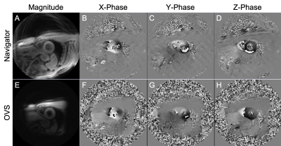
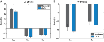
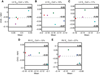
Total imaging time, including scouts and localizers, was 25±3 min (OVS) vs 47±8 min (navigator) (p<0.01). One subject had poor image quality and was excluded from the analysis. Magnitude and phase-encoded images for both sequences from a representative subject are shown in Figure 1. SNR of the magnitude images was high in both cases (mean±SD 58±10 Navigator vs 55±8 OVS; p=0.56). Global radial, circumferential, and longitudinal strains in the LV (Fig. 2A) and RV (Fig. 2B) were comparable between the two techniques. None of the differences in measured strains for either ventricle was statistically significant. Bland-Altman analysis and mean CoV showed good agreement for LV normal radial (CoV 12%), circumferential (10%), and longitudinal (11%) strains (Fig. 3A-C). Calculated RV strains were somewhat less consistent across protocols than those in the LV (CoV 12% circumferential, 27% longitudinal) (Fig. 3D-F).Discussion
DENSE, applied with OVS at 1.5T, is capable of measuring biventricular myocardial strain in a series of reasonable breath-holds. Such protocol developments may help to reduce the barriers to broader research applications and eventual clinical utilization, since the application is similar to other current standard cardiac MRI acquisitions.8A noteworthy limitation of this pilot study is the low power due to the small sample size. Further study with a larger, more heterogeneous sample will help to validate whether these methods give rise to equivalent strains. Differing field strengths and scan parameters may have an impact on strain agreement. However, other work has shown good agreement between similar sequences at these field strengths.9 As stated, RV strains were somewhat less consistent across protocols than those from the LV, suggesting the need for additional refinement of either the acquisition protocols and/or post-processing techniques. For this work, we assumed that the navigator-based data were more accurate due to previously demonstrated reproducibility characteristics. However, a separate validation of computed strains, e.g. from tagged MRI, may help to clarify the source of observed differences. Follow-up studies with such validation and more comprehensive inter-test reproducibility analysis will help address these limitations.
Conclusion
The acquisition of 3D biventricular myocardial strains during a breath-hold is feasible using 3D-encoded DENSE with OVS. While additional work is needed to validate the accuracy and reproducibility of myocardial strains derived from this method, these initial results suggest promising potential for a more generalizable and clinically feasible application of DENSE acquisitions for measuring 3D biventricular strain.Acknowledgements
This work was supported by the National Heart, Lung, and Blood Institute of the NIH under Award Number R01HL141901. The content is solely the responsibility of the authors and does not necessarily represent the official views of the NIH.References
1. Stanton T, Leano R, Marwick TH. Prediction of all-cause mortality from global longitudinal speckle strain: comparison with ejection fraction and wall motion scoring. Circ. Cardiovasc. Imaging 2009;2:356–64.
2. Kovács A, Lakatos B, Tokodi M, Merkely B. Right ventricular mechanical pattern in health and disease: beyond longitudinal shortening. Heart Fail. Rev. 2019.
3. Nagao M, Yamasaki Y, Yonezawa M, et al. Interventricular Dyssynchrony Using Tagging Magnetic Resonance Imaging Predicts Right Ventricular Dysfunction in Adult Congenital Heart Disease. Congenit. Heart Dis. 10:271–80.
4. Suever JD, Wehner GJ, Jing L, et al. Right Ventricular Strain, Torsion, and Dyssynchrony in Healthy Subjects Using 3D Spiral Cine DENSE Magnetic Resonance Imaging. IEEE Trans. Med. Imaging 2017;36:1076–1085.
5. Scott AD, Tayal U, Nielles-Vallespin S, et al. Accelerating cine DENSE using a zonal excitation. J. Cardiovasc. Magn. Reson. 2016;18.
6. Tayal U, Wage R, Ferreira PF, et al. The feasibility of a novel limited field of view spiral cine DENSE sequence to assess myocardial strain in dilated cardiomyopathy. Magn. Reson. Mater. Physics, Biol. Med. 2019;32:317–329.
7. Gilliam AD, Suever JD. DENSEanalysis. 2016. [Online]. Available: https://github.com/denseanalysis/denseanalysis
8. Kramer CM, Barkhausen J, Flamm SD, Kim RJ, Nagel E. Standardized cardiovascular magnetic resonance (CMR) protocols. 2013 update. J. Cardiovasc. Magn. Reson. 2013;15.
9. Wehner GJ, Suever JD, Haggerty CM, et al. Validation of in vivo 2D displacements from spiral cine DENSE at 3T. J. Cardiovasc. Magn. Reson. 2015;17:1–11.
Figures


