2188
Beyond SPAMM-PAV: a Phase Complementary SPAMM (PCSPAMM) acquisition to quantify simultaneously tissue motion and flow velocity1Biomedical Imaging Center, Pontificia Universidad Católica de Chile, Santiago, Chile, 2Millenium Nucleus for Cardiovascular Magnetic Resonance, Santiago, Chile, 3Department of Electrical Engineering, Pontificia Universidad Católica de Chile, Santiago, Chile, 4Department of Radiology, Pontificia Universidad Católica de Chile, Santiago, Chile
Synopsis
In this work, we introduce the Phase Complementary Spatial Modulation of Magnetization (PCSPAMM) acquisition sequence, designed to measure the tissue motion simultaneously with the flow velocity. With a pair of complementary acquisitions, PCSPAMM allows the estimation of a CSPAMM and a Phase-Contrast (PC) MR image, leading to improved tagging contrast and artifacts-free PC images at any cardiac phase.
Introduction
The cardiac function can be assessed using regional and simultaneous measurements of motion and velocities in the left-ventricle1. Previously, SPAMM EGG's2 and SPAMM-PAV3 have demonstrated to be useful techniques to obtain temporally correlated measures of both motion and velocity by combining a tagged MR acquisition with Phase-Contrast (PC) MRI. However, both approaches do not allow the measurement of early-systolic velocities and late-diastolic motion. In this work a new technique is presented, which combines a CSPAMM acquisition4 with PC, allowing the measurement of both motion and velocity across the whole cardiac cycle.Methods
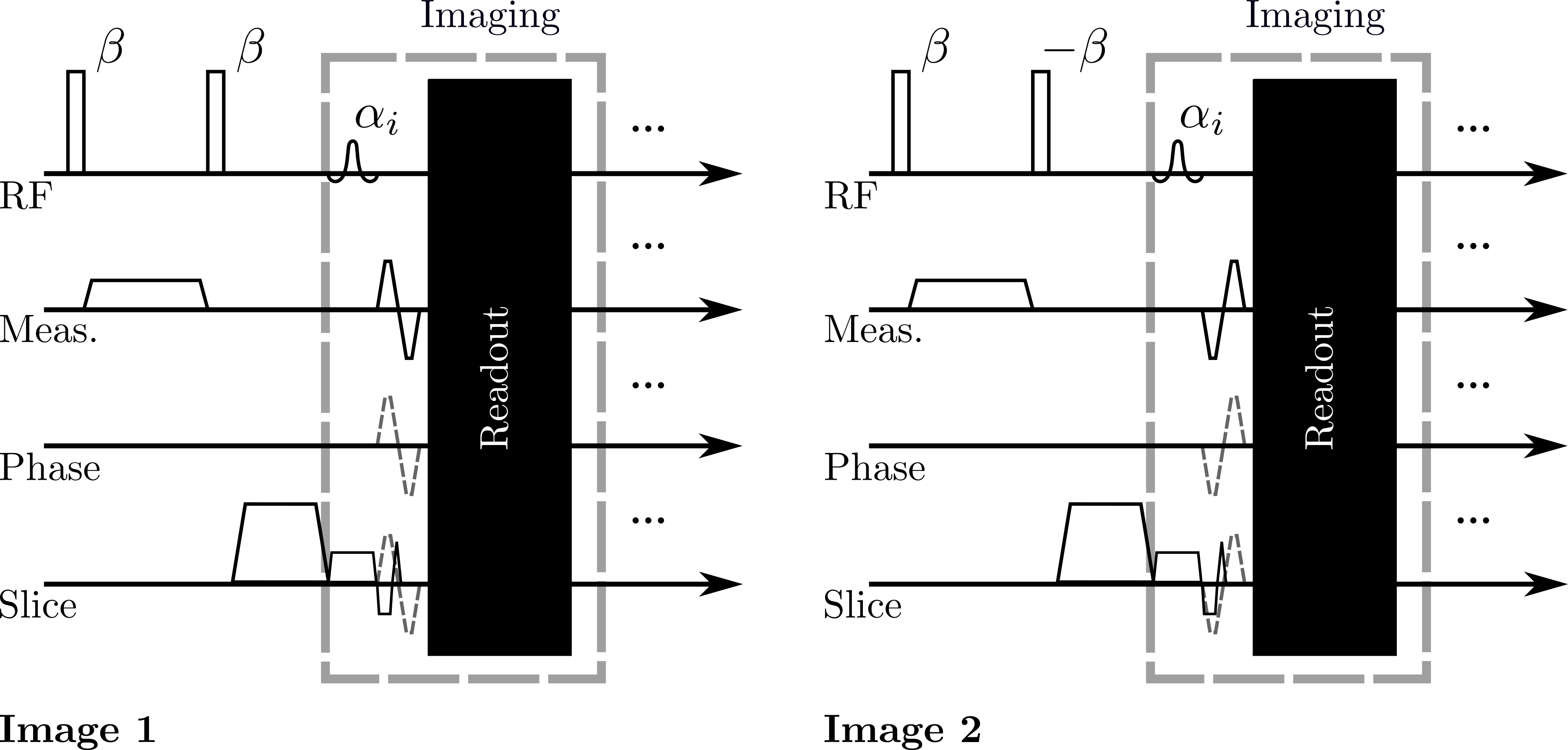
Similarly to SPAMM-PAV, a 1-1 CSPAMM acquisition was combined with PC-MRI by adding a bipolar gradient with alternate polarities to each SPAMM acquisition (see Figure 1). Two images containing information of the position and velocity of the tissue are measured, whose intensities are given by:$$
\begin{alignat}{2}
I_1 &= \left[a(\boldsymbol{p},T1(\boldsymbol{p}),t)+b(\boldsymbol{p},T1(\boldsymbol{p}),t)\cos(\phi_x(\boldsymbol{p},t))\right]e^{\boldsymbol{i}\phi_{in}(\boldsymbol{p})}e^{-\boldsymbol{i}\phi_v(\boldsymbol{p},t)} \\
I_2 &= \left[a(\boldsymbol{p},T1(\boldsymbol{p}),t)-b(\boldsymbol{p},T1(\boldsymbol{p}),t)\cos(\phi_x(\boldsymbol{p},t))\right]e^{\boldsymbol{i}\phi_{in}(\boldsymbol{p})}e^{\boldsymbol{i}\phi_v(\boldsymbol{p},t)}
\end{alignat}
$$
where $$$a$$$ and $$$b$$$ are increasing and decreasing functions depending on the position of the underlying tissue $$$\boldsymbol{p}$$$, the relaxation time $$$T1$$$, and the acquisition time $$$t$$$. In the previous expressions, $$$\cos(\phi_x)$$$ denotes the sinusoidal tagging pattern, $$$\phi_{in}$$$ the phase induced by field inhomogeneities, and $$$\phi_v$$$ the phase induced by the bipolar gradients.From the expressions given in the Equation 1, $$$\phi_{in}$$$ and $$$\phi_v$$$ are estimated using the following expressions:
$$
\begin{alignat}{2}
\phi_{in} &= \angle(I_1 + I_2) \\
\phi_{v} &= \angle(I_1e^{-\boldsymbol{i}\phi_{in}} + \text{conj}(I_2e^{-\boldsymbol{i}\phi_{in}})) \end{alignat}
$$
With both expressions estimated using the previous equations the images $$$I_1$$$ and $$$I_2$$$ are phase-corrected, allowing the generation of a CSPAMM image as described by Fischer et al4.
In this work we present a preliminary result consisting of a 4-chambers view of a healthy volunteer acquired in a 1.5T Achieva MR scanner (Philips, Best, Netherlands). We acquired two sets of PCSPAMM images allowing the obtention of tagged images in the read-out and phase directions and velocity encoded images in the phase and through-plane directions.
Results
In Figure 2 a comparison between the estimated CSPAMM image obtained with our proposal and the respective Magnitude Image CSPAMM Reconstruction (MICSR)5 is presented. As MICSR is a magnitude reconstruction, the phase of the acquisition did not distort the tag lines. In the case of the estimated CSPAMM image, the tagging pattern is similar to the obtained with MICSR, which means that the phase correction proposed in Equation 2 works properly.The Figure 3 shows the PC images obtained with our technique and a SPAMM-PAV-like reconstruction at the earliest systolic cardiac phase. The Figure 4 shows the mean velocities estimated using a SPAMM-PAV-like reconstruction and our proposal through the whole cardiac cycle. The mean values were calculated on a ROI positioned near the mitral valve (see Figure 3). The estimated velocity with PCSPAMM was close to the SPAMM-PAV results, giving mean peak velocity magnitudes of $$$57\pm10$$$ and $$$55\pm11$$$ cm/s respectively.
Discusion
The proposed technique allows the estimation of a PC and a CSPAMM using two complementary acquisitions for each encoding direction. The estimated PC was similar to the SPAMM-PAV estimation, but with our technique less artifacts were found at early diastolic cardiac phases (see Figure 3). In the case of the CSPAMM image, the tag persistence is improved as the DC spectral component is removed, allowing the estimation of motion through the whole cardiac cycle.Conclusion
We proposed a new acquisition technique that mix a CSPAMM acquisition with Phase-Contrast which allows the simultaneous estimation of motion and velocities through the whole cardiac cycle.Acknowledgements
Authors thankfully acknowledge the financial support of the CONICYT Ph. D. Scholarship 21170592, the project CONICYT-FONDECYT 1181057, and the Millennium Science Initiative of the Ministry of Economy, Development, and Tourism, grant Nucleus for Cardiovascular Magnetic Resonance.References
[1] Ziheng Zhang et al. Assessment of left ventricular 2D flow pathlines during early diastole using spatial modulation of magnetization with polarity alternating velocity encoding: A study in normal volunteers and canine animals with myocardial infarction. Magnetic Resonance in Medicine, 70(3):766–775, 9 2013.
[2] Smita Sampath, June H. Kim, Robert J. Lederman, and Elliot R. McVeigh. Simultaneous imaging of myocardial motion and chamber blood flow with SPAMM n’ EGGS (Spatial Modulation of Magnetization with Encoded Gradients for Gauging Speed). Journal of Magnetic Resonance Imaging, 27(4):809–817, 2008.
[3] Ziheng Zhang et al. Assessment of early diastolic strain-velocity temporal relationships using spatial modulation of magnetization with polarity alternating velocity encoding (SPAMM-PAV). Magnetic Resonance in Medicine, 66(6):1627–1638, 2011.
[4] S. E. Fischer et al. Improved myocardial tagging contrast. Magnetic Resonance in Medicine, 30(2):191–200, 8 1993.
[5] Moriel NessAiver et al. Magnitude image CSPAMM reconstruction (MICSR). Magnetic Resonance in Medicine, 50(2):331–342, 2003.
Figures