2183
Whole Heart High-Order B0 Shimming at 3T Using a UNIfied Coil (UNIC) for RF receive and shimming1Cedars-Sinai Medical Center, Los Angeles, CA, United States
Synopsis
B0-field inhomogeneity caused by tissue-air interface has been a long standing challenge in high field(>=3T) cardiac MRI. Although high-order shimming methods has been proposed in recent publications using surface shimming coils, the shimming capability in deep tissue, such as the heart, is still limited due to the restrained current amplitudes and the geometry constraints of the receiving loops. In this study, we developed a cardiac high-order shimming coil based on a novel coil decoupling design to overcome the aforementioned limitations. We demonstrated the developed coil can successfully reduce B0 field variation by 50% in the whole-heart in healthy human volunteers.
Introduction
Main field (B0) inhomogeneity has been a long standing challenge in high field(>=3T) cardiac MRI. The tissue-air interface between heart and lung induces strong local B0 variation, which is proportional to the scanner field strength. This effect degrades imaging capabilities of many important CMR sequences that are sensitive to off resonance, such as bSSFP, EPI readouts and fat saturation and T2 prep pre-pulses. It also diminish the SNR and spectral benefit from increased field strength and restrict CMR applications in high field scanners. Although state of the art scanners provide the capability of the 2nd spherical harmonic shimming, it often fell short when correcting the high order local field variations. In the past few years, RF coil arrays that integrate shimming currents into receiving RF loops were developed by us and other groups to enable high order local B0 shimming [1-3]. Although promising results has been presented in neurological applications, the shimming capability in deep tissue, such as the heart, is limited due to the restrained current amplitudes and the geometry constraints of the receiving loops. In this study, we use a novel technique we proposed recently, named unified coil (UNIC) [4,5]. UNIC overcomes the aforementioned limitations by decoupling the shimming and reception loops using a figure-8 configuration and by enabling multiple-turn shim loops to dramatically increase the shim field strength (Hardware design details will be discussed in an educational course on Multi-Coil B0 Field Modelling & Systems, ISMRM 2020). This allows shimming loops to be placed in close peripheral of the receiving coils and thus enable strong local shimming capacity. We tested the method in healthy human volunteers at a 3T clinical scanner.Methods
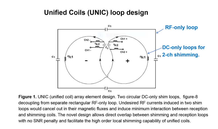
A unified coil was constructed with 12 RF receive and 42 shimming channels with 2-turn shim loops in each shim channel. The circuit design is depicted in figure 1. The constructed coil was placed on the subject’s chest wall to maximized SNR and shimming efficiency. Scans were performed in healthy human subjects (N=10) under breath holds and ECG gating. B0 field maps and bSSFP cine images with extended TR (TR= 6ms) were acquired to test the shimming efficacy. The field homogeneity and bSSFP image qualities were measured and compared with and without activating the UNIC shimming. Images were acquired After localization scans, whole-heart shimming, and scouting to determine the appropriate center frequency, breath-held, flow-compensated bSSFP acquisitions were prescribed in multiple planes along short-axis orientation with following scan parameters: TR = 6ms, flip angle = 50°, imaging resolution = 1.9 x 2.5 x 6 mm3, temporal resolution = 42 ms, readout bandwidth = 550 Hz;Results
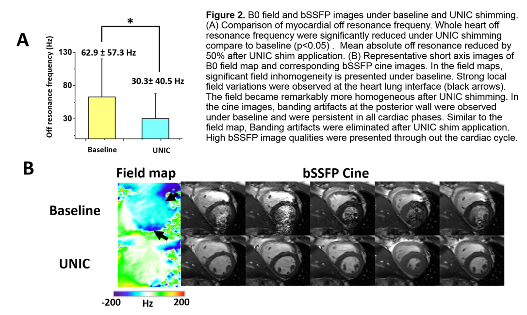
B0 Field and image qualities under baseline and UNIC shimming were compared in figure 2. Panel A presents the quantitative comparison of the myocardial off-resonance frequency with and without UNIC shimming. Off resonance frequency from each slice were measured. Both absolute mean and standard deviation of the myocardial off-resonance frequency were significantly reduced (62.9 ± 57.3 Hz vs 30.3± 40.5 Hz; p<0.05) after UNIC shimming. A set of representative short-axis images acquired with and without UNIC shimming is shown in panel B. Field maps and bSSFP cine images acquired at the matching slice position are presented. Significant field inhomogeneity is demonstrated at the heart-lung interface before UNIC shim application (black arrows). Banding artifacts at the posterior wall were observed in cine images and strongly degrade the image qualities throughout the cardiac cycle. After UNIC shim application, B0 field became remarkably more homogeneous in the myocardium and the banding artifacts were eliminated in the bSSFP cine images.Conclusion
To our best knowledge, this is the first study to perform local cardiac shimming in a clinical high field scanner. UNIC coils successfully reduce off-resonance frequency by 50% in the heart at 3T. The field improvement opens up the opportunity for reliable CMR in high field scanners and can potentially unleash the full SNR and spectral benefit for CMR applications.Acknowledgements
Thanks to Fei Han, Bernd Stoeckel, Fraser Robb and Miguel Navarro for their support.References
1. Han H et al. MRM 2013;70:241-247.
2. Truong TK et al. Neuroimage 2014;103:235-240.
3.Stockmann JP, Witzel T, Keil B, et al. A 32-channel combined RF and B0 shim array for 3T brain imaging. Magn Reson Med 2016;75:441-451.
4. Hui Han, John Stager, Hsin-Jung Yang, Sizhe Guo, Zhuoqi Li, and Debiao Li. Unified Coils (UNIC) for Parallel Imaging and B0 Shimming. 2016 Gordon Research Conference, In Vivo Magnetic Resonance, 'MRI Inside-Out and Outside-In: Innovative Technologies, Unmet Needs and New Opportunities'. Proctor Academy, Andover, NH. July 17-22, 2016.
5. H. Han, J. Stager, W. Cao, Z. Li, J. Cho, D. Zhou, Y. Wang, D. Li. Unified Coils (UNIC) for Parallel Imaging and B0 Shimming. European Society for Magnetic Resonance in Medicine and Biology (ESMRMB). ESMRMB 2016 Congress. 33rd Annual Scientific Meeting, Vienna/AT, September 29 - October 1, 2016. (Oral presentation).
Figures