2176
Auto-calibrated simultaneous multi-slice cardiac bSSFP Cine MRI using gradient temporal modulation
Yuan Zheng1, Lele Zhao2, Zhongqi Zhang2, Zhehao Zhang2, Xingxian Shou1, Haojie Li3, Jian Xu1, Weiguo Zhang1, Lu Huang3, and Liming Xia3
1UIH America, Houston, TX, United States, 2United Imaging Healthcare, Shanghai, China, 3Tongji Hospital, Wuhan, China
1UIH America, Houston, TX, United States, 2United Imaging Healthcare, Shanghai, China, 3Tongji Hospital, Wuhan, China
Synopsis
Simultaneous multi-slice (SMS) imaging is an efficient MRI technique that better preserves SNR than conventional parallel imaging (PI). However, single-band images usually need to be acquired to provide references for slice-unaliasing, causing an untrivial scan time overhead in some scenarios. We extended a recently reported self-calibrated SMS imaging strategy based on RF phase modulation, and propose using gradient temporal modulation to achieve auto-calibrated SMS bSSFP, which avoids the bSSFP banding shift and is preferred in cardiac Cine due to its contrast and intrinsically high SNR. In-plane PI can also be incorporated with shared auto-calibration.
Introduction
Simultaneous Multi-Slice (SMS) imaging is a technique that improves the time efficiency of MRI pulse sequences by exciting and imaging multiple slices simultaneously1. However, single-band images usually need to be acquired separately to provide references for the slice-unaliasing task2, causing an untrivial scan time overhead in some scenarios, especially in certain cardiac imaging applications that require breath-holding (BH). Recently an auto-calibrated SMS CAIPIRINHA imaging technique which eliminated the need for extra reference scans was reported3. An additional RF phase modulation was applied along the temporal dimension, enabling extraction of single-band references from the multiband (MB) data. However, RF phase modulations may introduce artifacts in balanced steady-state free precession (bSSFP) sequences, since spins are subjected to an additional phase each TR and the bSSFP banding artifacts would be shifted. In this abstract, we propose using gradient modulation to achieve the temporal phase encoding required for auto-calibrated SMS. Since TR-TR signal evolution is not affected, the bSSFP bandings remain unchanged. In-plane parallel imaging (PI) can also be integrated into this framework and share the auto-calibration property. We implemented this technique in bSSFP cardiac Cine sequences and demonstrated its advantages in clinical applications.Theory
Phase modulation of the proposed technique is achieved by modifying the rephrasing and prephasing gradient lobes4, as shown in Fig.1. The gradient pair is modified such that a desired phase shift is added to the signal before ADC but cancelled out afterwards. Therefore, the net gradient moment remains zero and the TR-TR spin evolution is unchanged. For auto-calibrated SMS CAIPIRINHA imaging, the gradient induced phases serve two purposes: within a frame they cause controlled aliasing; between frames they create controlled phase offsets of the collapsed slices, allowing extraction of single-band reference images. The phase modulation table and image reconstruction procedure for MB = 2 are shown in Fig.2. Taking slice 1 as reference, the linear phase modulation causes an FOV/2 shift of slice 2 with a phase offset varying by π between even and odd frames. The reference images can be extracted by summation and subtraction of adjacent frames. Although the reference images are slice unaliased, they are of lower temporal resolution because different time frames are mixed. The reference images are then used for kernel calibration to unalias single-frame MB images using slice-GRAPPA5.The phase modulation table and steps for extracting single-band reference images for MB = 2 and PI = 2 are shown in Fig.3. Frame 1, 2 samples odd PE lines, while Frame 3, 4 samples even PE lines, etc, in a manner similar to temporal GRAPPA6. The phase offset of slice 2 alternates by π between frames. Four frames are needed to extract the single-band references. Taking the first four frames for example, frames 1, 2 and 3, 4 are first combined respectively to separate the collapsed slices, leaving single-band images with in-plane ghosting. The ghosting patterns are different depending on whether even (frame 3, 4) or odd (frame 1, 2) lines are sampled. By combining them, ghost-free images are obtained, which can be used as reference images for reconstructing single-frame images from the MB data, using slice-GRAPPA and GRAPPA algorithms.
Methods
The proposed auto-calibrated SMS bSSFP Cine was tested on a healthy volunteer on a 3.0 T scanner (uMR780, United Imaging Healthcare, Shanghai, China), with 40 receive channels (a 24-channel cardiac coil and a 16-channel spine coil). Imaging parameters include: FOV = 360×300 mm, matrix = 224×224, thickness = 8 mm, 2 slices were imaged with gap = 40 mm, bandwidth = 1200 Hz/pixel, FA = 45°, TR/TE = 3.1/1.4 ms, frame # = 23 per heartbeat, views per frame = 13. Four scans were performed with no acceleration, PI (2), MB (2), and MB (2) + PI (2). The number of heart beats needed were: 34 for no acceleration; 17 for PI (2); 17 for MB (2) and 9 for MB (2) + PI (2). The no-acceleration scan was completed in two BHs, while all others were collected within a single BH.Results
Fig.4 compares images of the same cardiac phase acquired with different approaches. Heart structures were well delineated in all images. No obvious artifacts were observed, although a visible SNR drop was observed in the MB (2) + PI (2) image, which is expected due to undersampling of k-space and the g-factor penalty. Fig.5 zoomed in on the heart and shows 10 consecutive frames acquired with MB (2) and MB (2) + PI (2). Wall motion was well preserved in both Cine image series.Discussion and Conclusion
SMS is an SNR efficient MRI acceleration technique and is preferred over conventional PI in many applications. Based on a recently proposed auto-calibrated SMS imaging concept, we used gradient temporal modulation to achieve auto-calibrated SMS bSSFP, which avoids the bSSFP banding shift and is preferred in cardiac Cine due to its contrast and intrinsically high SNR. We also showed that in-plane PI can be incorporated with shared auto-calibration. The proposed technique was demonstrated on volunteers and achieved high-quality Cine images. Overall, our proposed technique can be used to collect SMS bSSFP images without dedicated reference scans and could be used to simplify the clinical scan work flow and reduce scan time.Acknowledgements
No acknowledgement found.References
- Barth M et al., Simultaneous multislice (SMS) imaging techniques, Magn Reson Med. 2016 Jan;75(1):63-81.
- Breuer FA, et al., Controlled aliasing in parallel imaging results in higher acceleration (CAIPIRINHA) for multi-slice imaging, Magn Reson Med. 2005 Mar;53(3):684-91.
- Ferrazzi G, et al., Autocalibrated multiband CAIPIRINHA with through-time encoding: Proof of principle and application to cardiac tissue phase mapping, Magn Reson Med. 2019 Feb;81(2):1016-1030.
- Zheng Y,et al., Comparison of Methods for Simultaneous Multi-Slice Balanced SSFP Imaging, ISMRM 2017:1271.
- Setsompop K, et al., Blipped-controlled aliasing in parallel imaging for simultaneous multislice echo planar imaging with reduced g-factor penalty. Magn Reson Med. 2012 May;67(5):1210-24.
- Breuer FA et al., Dynamic autocalibrated parallel imaging using temporal GRAPPA (TGRAPPA), Magn Reson Med. 2005 Apr;53(4):981-5.
Figures
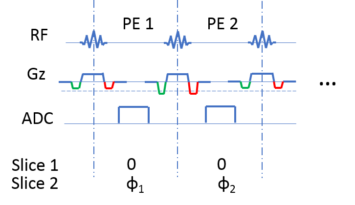
MR signal phase can be
modulated by gradients. A phase difference ϕ can be introduced between slices
by a gradient moment Mz=ϕ/γd,
where γ is the gyromagnetic ratio and d is the distance between slices. In
SMS-bSSFP sequences, the rephrasing (red) and prephasing (green) gradient lobes
are both modified while keeping the total gradient balanced, i.e., with no net
zeroth moment.
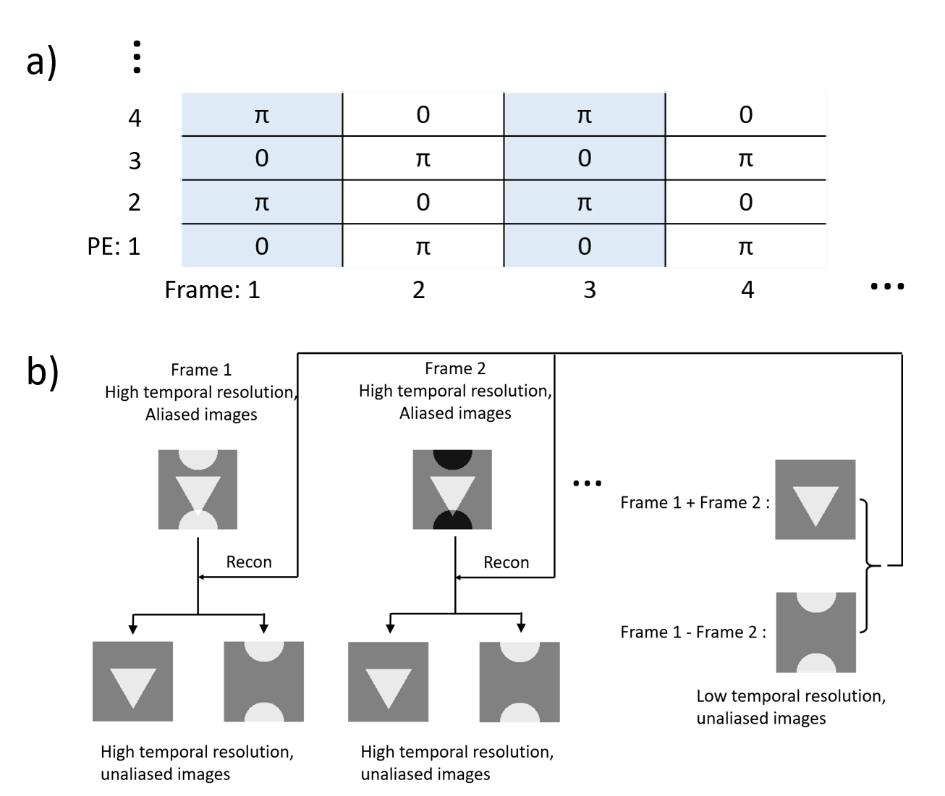
a), the phase modulation
table for MB = 2. Phase differences between the two slices for each PE line
are listed. The linear phase progression in each frame induces FOV/2 controlled
aliasing, while the π phase difference between even and odd frames enables
separation of the collapsed slices. b), in image reconstruction, single-band references are first calculated by properly mixing the frames, and then used to
unalias the single-frame collapsed slices.
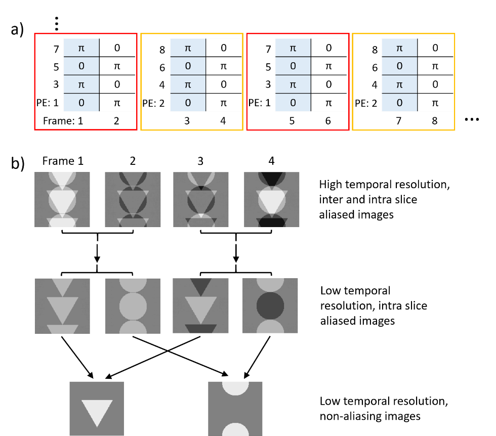
a), the phase modulation table for MB (2) + PI (2). Phase differences
between the two slices for each PE line are listed. In all frames there is a π
phase progression between subsequently acquired PE lines, and there is a π
phase difference between even and odd frames. Besides, only odd PE lines are
sampled for frames 1, 2, 5, 6, etc. (red box), while only even PE lines are
sampled for frames 3, 4, 7, 8, etc. (orange box). b), unaliased single-slice
images can be extracted by properly combining four adjacent frames.
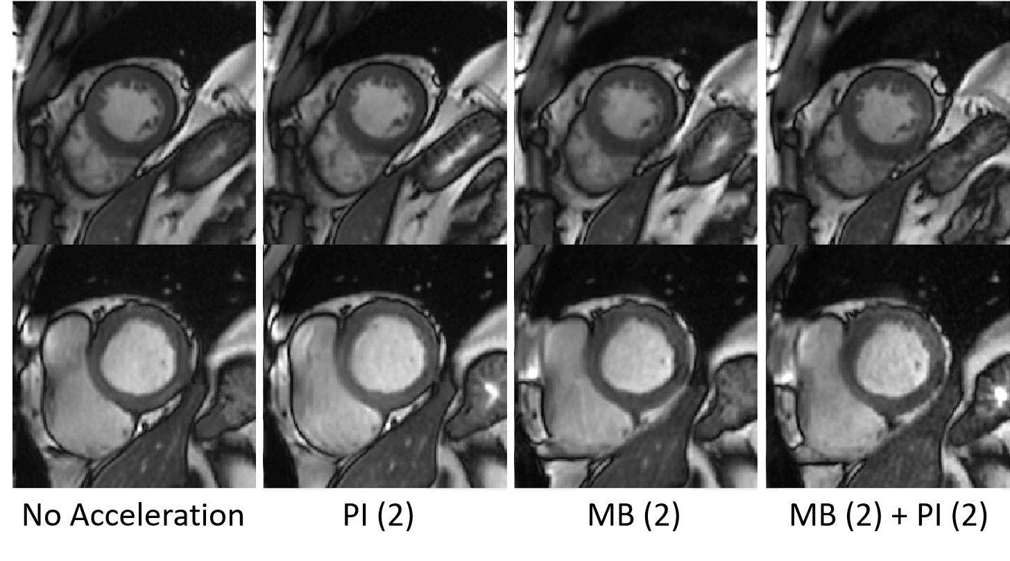
Comparison of images acquired with different acceleration parameters. Without
SMS the two slices were acquired separately (columns 1 and 2); With SMS the slices were
collected simultaneously (columns 3 and 4). Cardiac morphology was well delineated in
all images.
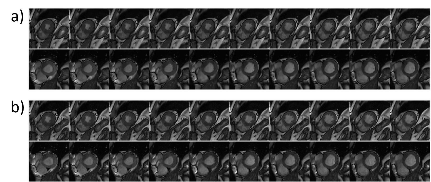
Ten consecutive frames from systole to diastole phase. Two slices were imaged simultaneously with MB
(2) only (a) and MB (2) + PI (2) (b) acquisitions. The temporal
resolution of each frame was 3.1 ms × 13 views = 39 ms. Wall motion was well preserved in both series.