2171
Accelerated-x4 Balanced-SSFP Cardiac Cine 2D/3D DENSE with Phase-Cycled Encoding for Improved Performance1MRI Research Center, Auburn University, Auburn University, AL, United States
Synopsis
Cine frame-rate quantification of myocardial strain has been previously demonstrated with 2D echo-planar and 2D/3D spiral sequence versions of Displacement Encoding with Stimulated Echoes (DENSE). While these non-conventional methods allow fast-acquisition, they are plagued with off-resonance limitations that has hindered their integration into mainstream cardiac MRI application. Here we present a more conventional, accelerated-x4, balanced SSFP (bSSFP) version of Cardiac Cine 2D/3D DENSE with improved phase-cycled encoding for effective artifact suppression. In vivo human scans at 3T demonstrated good agreement of myocardial radial (Err), circumferential (Ecc) and longitudinal (Ell) strains as reported in previous literature along with lower artifact degradation compared to standard complementary-encoded DENSE.
Introduction
Quantification of myocardial strain has been previously demonstrated with echo-planar (2D) and spiral (2D&3D) sequence versions of Displacement Encoding with Stimulated Echoes (DENSE)1-3. However, these non-conventional acquisition methods, with their sensitivities to off-resonance, has hindered their integration into mainstream cardiac MRI (CMR) application. Balanced Steady-State Free Precession (bSSFP) that’s known for high SNR and versatility is well favored in CMR. Past efforts to integrate displacement encoding methods into bSSFP has shown limited success4, 5. Here we present a bSSFP version of Cardiac Cine 2D/3D-DENSE that incorporates high x4 GRAPPA acceleration and 3-Point Phase-Cycled Encoding (PhsCyc) that eliminates the undesired T1 recovery and encoded anti-echo signals. PhsCyc has been demonstrated in Spiral DENSE3 at 1.5T, but here we apply it in bSSFP on human subjects at 3T.Methods
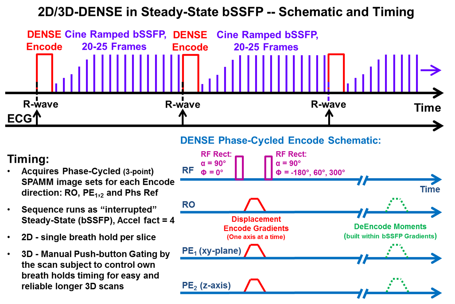
Fig-1 illustrates the PhsCyc bSSFP Cine DENSE sequence that runs as ECG-triggered interrupted steady-state to acquire data on every heartbeat. The PhsCyc DENSE encoding applies a 3-point phase rotation to the second RF pulse that encodes the images for subsequent DeEncode to eliminate the T1 and anti-echo (AE) signal components. The PhsCyc encoding can be applied to in-plane, or through-plane encoding. The Encode/DeEncoding process consists of three linear equations to solve for three unknowns, therefore the DeEncode process is simply the inverse of the Encode matrix. The cine bSSFP readout is customized with ramped start-up flip angle for fast approach to steady-state and modified gradients to incorporate the DENSE de-encode moments while maintaining gradient moment nulling in all axes. The end of cardiac R-R cycle can incorporate either α/2 magnetization restore method, or only spoiler gradients before the next triggered DENSE encode. For each 3D partition, the DENSE serially acquires 12 PhsCyc image sets including encoded readout (x-axis), encoded phase-encode (y-axis), encoded through-plane (z-axis), and non-encoded phase-reference directions. For 2D, only 9 images sets are required and requires only one breath hold. For each encoded axis, the bSSFP readout module integrates the necessary de-encode moments into its relevant gradients. Imaging parameters for bSSFP 3D-DENSE: FOV = 260x240 mm, Partitions = 16, Partition thickness = 5 mm, Matrix = 112x90, Pixel size = 2.5x2.5x5 mm, Bandwidth = 1116 Hz/Pixel, GRAPPA acceleration = x4, Flip angle = 20°, Cine frames = 20, Averages = 1, DENSE in-plane/through-plane encode = 0.12/0.08 cyc/mm. 3D-DENSE scans placed the 8-10 partitions at mid-Left Ventricle (LV) with the partition “slices” as cardiac short-axis (SA) direction. All image and strain analysis were performed offline using customized Matlab programs (Mathworks, Natick, MA). Myocardial mid-LV SA, six sector and global average strain values for radial (Err), circumferential (Ecc), and longitudinal (Ell) directions were calculated and presented. Importantly, similar to navigator gating, our sequence employed a manual push-button gating method. The subject being scanned would press and hold the gating button during exhaled breath-hold (allowing acquisition) then release the button before and during breathing (non-acquisition steady-state). The button process is repeated on the subject’s next breath-hold and repeated as necessary for the duration of the scan. Four healthy human subjects, 22-52yo, 1 female, with informed consent, were scanned in a 3T Verio scanner (Siemens, Erlangen, Germany) with a 32-chan anterior/posterior RF coil array (Invivo, Gainesville, Florida).Results
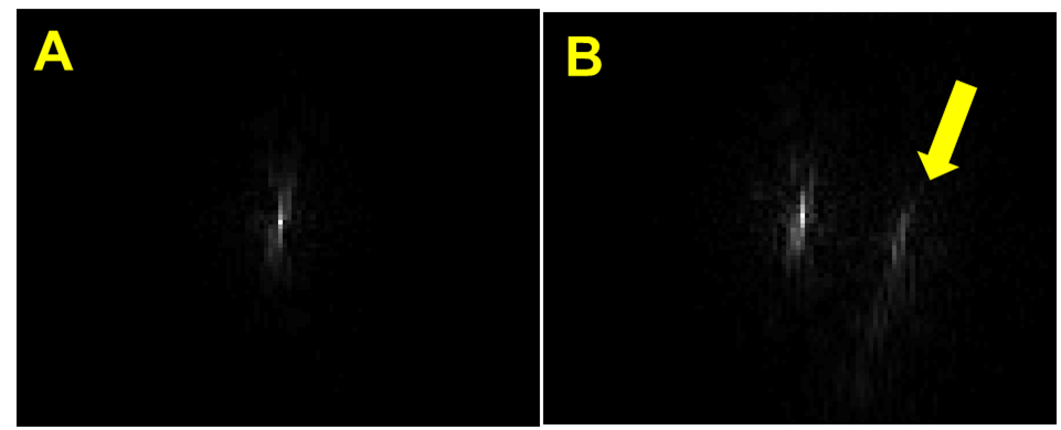
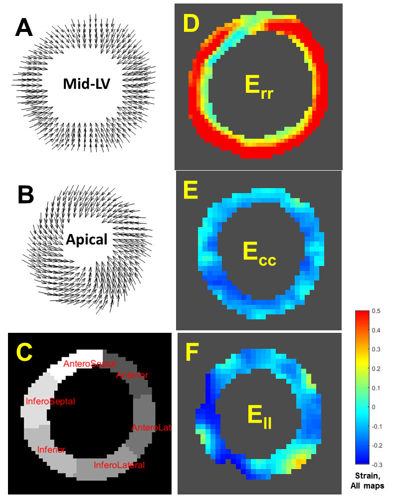
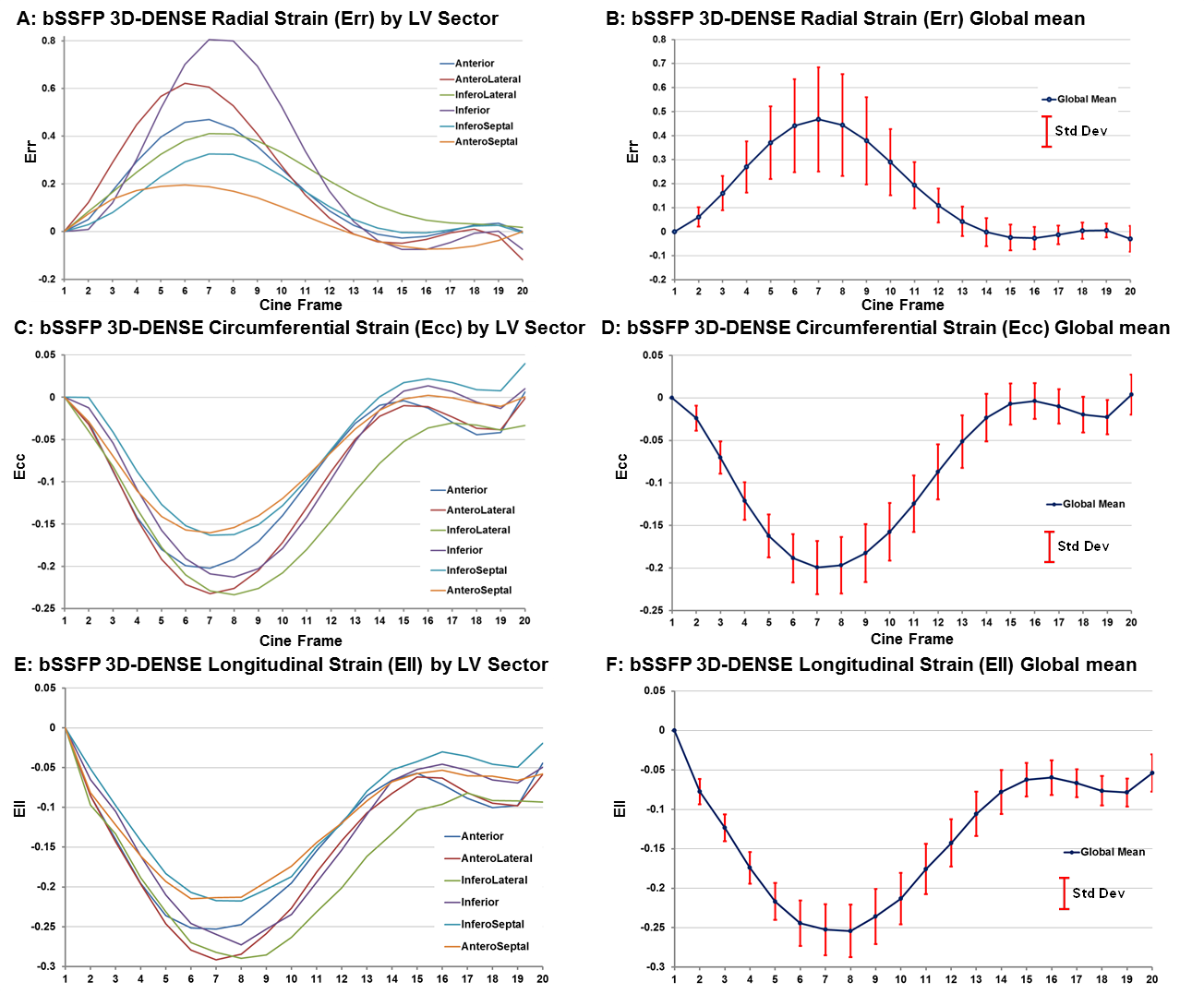
From all subjects, bSSFP DENSE provided displacement encoded and reference image sets suitable for strain analysis. The manual push-button gating method proved very easy and effective for acquiring long (10-15 minute) multi-breath-hold scans. The PhsCyc was highly effective at removing the T1 and AE signals compared to standard CSPAMM as shown in Fig-2. Figs-3&4 present representative DENSE image and strain analysis results at mid-LV. Fig-2 shows good 2D in-plane displacement field patterns for mid-LV and apical slices. Fig-3 presents strain maps for the three Err, Ecc, and Ell directions. Fig-4 presents AHA-convention, six-sector strain and global mean strain graphs for the three Err, Ecc, and Ell directions for each cine frame. Subjects with slow heart-rates (RR > 1 sec) showed increased DENSE signal fading and subsequent noise in the late-diastole frames – as evident in the Fig-4 strain graphs The bSSFP DENSE showed good agreement per subject, however, variation was high across subjects with global mean Err = 0.38±0.30, Ecc = 0.19±0.21 and Ell = 0.23±0.22 (Mean±StdDev).Discussion
This accelerated bSSFP 3D-DENSE with PhsCyc for human 3D cardic strain quantification has performed well. The PhsCyc was effective enough that 2D DENSE scans were run without Through-plane encoding to eliminate the T1 and AE. The x4 acceleration with a long 3D scan gave good SNR (≈>50). The DENSE T1 fading was sometimes problematic, so work is needed to optimize the bSSFP flip angle method. The manual push-button gating works well with cooperative subjects, but impractical for actual medical patients.Conclusions
We developed and demonstrated an accelerated bSSFP Cine 2D/3D DENSE sequence with Phase-Cycled encoding for human 3T CMR application. Future work will improve on its overall performance.Acknowledgements
Special thank you for programmatic and volunteer support goes to Julie Rodiek, Julio Yanes, Adam Davila and Lily Strassberg.References
1. Aletras AH, Ding S, Balaban RS, Wen H. “DENSE: Displacement Encoding with Stimulated Echoes in Cardiac Functional MRI”, J Magn Reson. 1999; 137(1): 247–252.
2. Kim D, Gilson WD, Kramer CM, Epstein FH, “Myocardial Tissue Tracking with Two-dimensional Cine Displacement-encoded MR Imaging: Development and Initial Evaluation”, Radiology 2004; 230:862-871.
3. Zhong X, Spottiswoode BS, Meyer CH, Kramer CM, Epstein FH, “Imaging Three-Dimensional Myocardial Mechanics Using Navigator-gated Volumetric Spiral Cine DENSE MRI”, MRM 2010; 64(4):1089-1097.
4. Cowart EA, Gilson WD, Kramer CM, Epstein FH, “Imaging 3D Myocardial Motion with SSFP Cine DENSE”, Proc. Intl. Soc. Mag. Reson. Med. 11, 2004.
5. Kim D, Kellman P, “Improved cine displacement-encoded MRI using balanced steady-state free precession and time-adaptive sensitivity encoding parallel imaging at 3T”, NMR Biomed 2007; 20(6):591-601.
Figures