2169
An Efficient Sampling Strategy of Fast Multi-slice Cardiac Cine Imaging
Dongchan Kim1, Byungjai Kim2, Kyoung-Nam Kim3, Jun-Young Chung4, HyunWook Park2, and Yeji Han3
1Samsung Electronics, Seoul, Republic of Korea, 2Department of Electrical Engineering, Korea Advanced Institute of Science and Technology, Daejeon, Republic of Korea, 3Department of Biomedical Engineering, Gachon University, Incheon, Republic of Korea, 4Gachon Advanced Institute for Health Sciences and Technology, Gachon University, Incheon, Republic of Korea
1Samsung Electronics, Seoul, Republic of Korea, 2Department of Electrical Engineering, Korea Advanced Institute of Science and Technology, Daejeon, Republic of Korea, 3Department of Biomedical Engineering, Gachon University, Incheon, Republic of Korea, 4Gachon Advanced Institute for Health Sciences and Technology, Gachon University, Incheon, Republic of Korea
Synopsis
Balanced steady-state free precession (bSSFP) with electrocardiogram (ECG) triggering and breath-holding is a common MRI technique for examination of ventricular function. For whole-heart imaging, resting periods are often necessary between breath-holds because many patients find it difficult to perform successive breath-holds and this eventually prolongs the entire examination time. Although self-gating (SG) techniques can relieve the discomfort that comes from multiple breath-holds, the sampling efficiency is limited because the data from inspiration phases are discarded. In this work, we propose a new sampling strategy for efficient multi-slice cardiac cine imaging that acquires data with a reduced number of breath-holds.
Background
In clinical settings, cardiac magnetic resonance imaging (CMRI) is commonly performed with breath-holding, while synchronizing the data acquisition timing with the electrocardiogram (ECG) signals of external ECG devices for examination of ventricular function [1,2]. For whole-heart imaging, resting periods are often necessary between breath-holds because many patients find it difficult to perform successive breath-holds and this eventually prolongs the entire examination time. Although self-gating (SG) techniques can relieve the discomfort that comes from multiple breath-holds, the sampling efficiency is limited because the data from inspiration phases are discarded. In this work, we propose a new sampling strategy for efficient multi-slice cardiac cine imaging that acquires data during a single breath-hold, directly followed by a free-breathing scan without an interval.Methods
In the proposed method, data acquisition is continuously performed without a break during a single breath-hold and a subsequent free-breathing period, where the entire k-space is sampled to acquire multiple slices of images for cardiac cine MRI. In the breath-hold period, the k-space signals of the pre-defined low-frequency section are acquired to ensure the image quality, where the number of phase-encoding lines for low-frequency section (NLF) is determined by the repetition time (TR), the number of slices (Nslc), the number of cardiac phases (Nph), and the optimal breath-hold time (tBH) of each patient, as NLF= tBH/(TR×Nph×Nslc) (eq.1). For multiple slices, the central k-space signals from –NLF/2 to NLF/2 are repeatedly acquired during the breath-hold period to generate images of multiple cardiac phases (Fig.1). Then, the remaining high-frequency k-space region is filled during the free-breathing period and to generate the final multi-slice images using SPiRiT [3].Experiments were performed with five healthy volunteers (age: 29.8±4.2) using a 3.0 T MRI scanner (Siemens MAGNETOM Verio, Erlangen, Germany) under institutional review board approval. For the proposed method, the following parameters were used with the phase-based SG technique [4]: FOV = 256 × 256 mm, matrix size = 256 × 256, slice thickness = 8 mm, flip angle (FA) of SPGR sequence for SG = 45°, FA for SSFP-FID sequence for imaging = 17°, TR/TE = 6.9/2.0 ms, VENC = 120 cm/s, Nph = 20. tBH of 10 s was used based on the previous studies [5,6] to reconstruct six image slices and the free-breathing period of 3 min per each slice was used. To cover the whole heart region, twelve slices were acquired with two breath-holds. Then, we retrospectively subsampled the data acquired during the free-breathing period to analyze the image quality with respect to the scan time. To obtain the same amount of low-frequency k-space signals and the same number of total slices from patients with highly limited breath-holding capability, i.e., a short breath-hold period, the number of acquired slices during a single breath-hold needs to be reduced, requiring an increased number of breath-holds. To evaluate the performance of the proposed sampling scheme with respect to the duration of a single breath-hold, we also analyzed the quality of cine images reconstructed from data acquired during breath-holds of 0, 10, 20 s and free-breathing of one minute. For this experiment, the number of frequency-encoding lines with respect to different breath-hold periods were accordingly calculated using eq.1.
Results
Fig. 2 shows the images acquired with the proposed method (tBH = 10 s) and the phased-based SG methods using various imaging times, where the free-breathing periods varied according to the total imaging time. For imaging time of 3 min, both the proposed method and the phased-based SG method successfully generated cardiac cine images with satisfactory subjective image quality. Similarly, the images of the proposed method showed satisfactory image quality when the imaging times were reduced to 2 min and 1 min. However, the images of the phased-based SG method showed severe aliasing artifacts due to the missing k-space data in the low-frequency region. In addition, different breath-hold periods with a fixed free-breathing period of 1 min were used to demonstrate the effect of the low-frequency data acquired during the breath-hold periods. As demonstrated by Fig.3, the proposed method improved the quality of cine images according to the duration of the breath-holds because more amount of central k-space data could be obtained. In a practical situation, possible durations of breath-hold can vary according to the breath-holding capability of the patients. When the breath-holding duration is limited to less than 10 s, the acquisition time with free-breathing can be increased to longer than 1 min to obtain sufficient data for cine images with similar quality to those acquired with 20 s of breath-holding.Discussion and Conclusions
Compared with the phase-based SG technique, the proposed method significantly reduced the total imaging time, while preserving the image quality. Moreover, it can be also applied to various cardiac cine imaging methods. For example, the proposed imaging scheme can be applied to the conventional bSSFP cardiac cine imaging to reduce the required number of breath-holds by utilizing an additional 1 min of free-breathing period. In summary, the proposed method can perform cardiac cine MRI with reduced imaging time and reduced number of breath-holds. The experiment results show that the proposed method can be applied to the conventional cardiac cine techniques and maintains consistent image quality.Acknowledgements
No acknowledgement found.References
- Ginat DT, Fong MW, Tuttle DJ, Hobbs SK, Vyas RC. Cardiac imaging: Part 1, MR pulse sequences, imaging planes, and basic anatomy. Am J Roentgenol. 2011;197:808–15.
- Kramer CM, Barkhausen J, Flamm SD, Kim RJ, Nagel E. Standardized cardiovascular magnetic resonance (CMR) protocols 2013 update. J Cardiovasc Magn Reson. 2013;15:91.
- Lustig M, Pauly JM. SPIRiT: Iterative self-consistent parallel imaging reconstruction from arbitrary k-space. Magn Reson Med. 2010;64:457–71.
- Seo H, Kim D, Oh C, Park HW. Self-gated cardiac cine imaging using phase information. Magn Reson Med. 2017;77:1216–22.
- Liu J, Spincemaille P, Codella NCF, Nguyen TD, Prince MR, Wang Y. Respiratory and cardiac self-gated free-breathing cardiac CINE imaging with multiecho 3D hybrid radial SSFP acquisition. Magn Reson Med. 2010;63:1230–7.
- Larson AC, White RD, Laub G, McVeigh ER, Li D, Simonetti OP. Self-Gated Cardiac Cine MRI. Magn Reson Med. 2004;51:93–102.
Figures
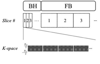
A schematic diagram of the proposed method for multi-slice cardiac cine imaging.
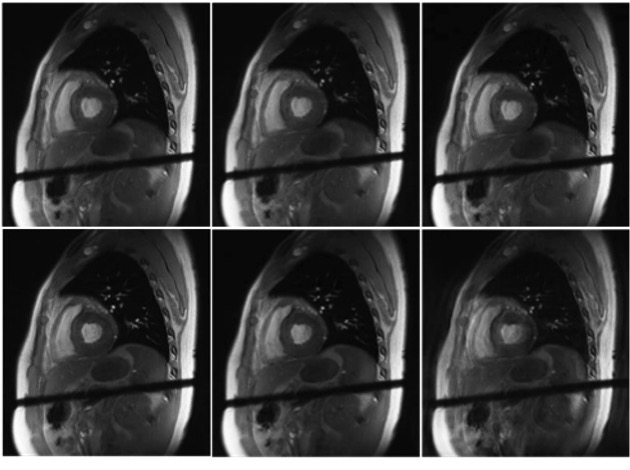
Cardiac images of the proposed method with tBH = 10 s (top row) and the free-breathing method (bottom row) acquired in 3 (left), 2 (middle), and 1 (right) minutes, respectively. The proposed method generated cine images of 20 cardiac phases with satisfactory image quality, while images acquired only with free-breathing showed motion-related artifacts.
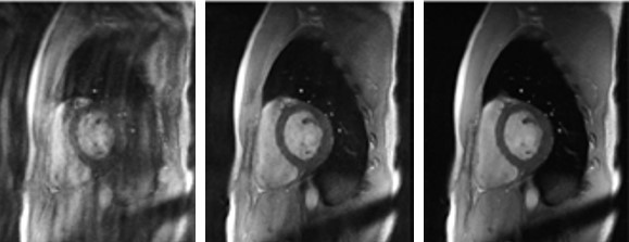
Comparison of cardiac images acquired with different breath-hold durations using the proposed method: no breath-hold (left), 10 s (middle), and 20 s (right). All datasets were acquired with a 1 min long free-breathing period.