2166
CMR Feature tracking: reproducibility in small animals1Core Facility Small Animal Imaging, Ulm University, ULM, Germany, 2Department of Cardiology, The Second Hospital of Shandong University, Jinan, China, 3Department of Internal Medicine II, Ulm University Medical Center, Ulm, Germany
Synopsis
Cardiac magnetic resonance imaging feature tracking (CMR-FT) is a novel non-invasive imaging technique and has been validated in clinical applications. However, there are only limited studies in small animal research. We performed biventricular strain assessment using CMR-FT in nexiline knock-out mice at rest and dobutamine stress to evaluate its feasibility and intra-observer reproducibility in preclinic research. Excellent intra-observer reproducibility could be observed for rest and stress left ventricular (LV) strain in general and right ventricular (RV) circumferential strain derived from short-axis views. An only fair reproducibility was observed for RV longitudinal strain under stress conditions derived from long-axis views.
Purpose
To investigate the feasibility and intra-observer reproducibility of CMR-FT in biventricular strain assessment in the mouse model.Introduction
Myocardial strain is a well validated parameter for evaluating myocardial performance. Since the introduction of myocardial tagging technique by Zerhouni in 19881, measurement of myocardial strain using CMR imaging became possible. However, the requirement for extra image acquisition sequences and time-consuming post process limits its application. Recently, feature tracking was introduced to calculate myocardial strain for global and segmental functional assessment2. As CMR-FT is a promising novel method for the quantification of myocardial strain from standard steady-state-free-precession (SSFP) images without prolonged post processing times, it has the potential for fast assessment of myocardial mechanics. Its clinical potential has been recently described3–5, and excellent inter and intra observer agreement and high inter-study reproducibility were reported. However, only limited studies were reported in preclinic research6. In this contribution, we perform CMR-FT in a congestive heart failure mouse model to investigate the feasibility and intra-observer reproducibility of biventricular strain assessment.Methods
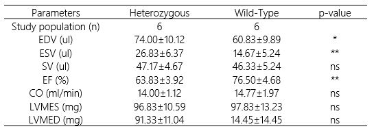
All measurements were performed on an 11.7 Tesla small animal MRI system (BioSpec 117/16, Bruker Biospin, Ettlingen, Germany) with a four-element thorax coil optimized for imaging the mouse heart (RAPID Biomedical, Rimpar, Germany). Reference functional data were acquired applying a conventional Cartesian self-gated sequence (IntraGate©, ParaVision 5.1, Bruker Biospin, Ettlingen,Germany)7. Slice planning was performed as suggested earlier7 ensuring high reproducibility of the image geometry. The number of short axis slices was adjusted to ensure full coverage of the left ventricle in end-diastole. Typical imaging parameters: echo/repetition time TE/TR = 1.0/5.75ms, flip angle α = 15°, matrix size = 256x256, in-plane resolution Δr = 0.1232mm2, slice thickness sD = 0.5mm, bandwidth bw = 150KHz, 20 phases per cardiac cycle, acquisition time TACQ = 42s per slice.The imaging protocol was performed in 12 constitutive nexiline (Nexn) knock-out (KO) mice, heterozygous (Het, N=6) and wild-type (WT, N=6). During scanning, the animals were kept at mild anaesthesia (respiratory rate > 60 cpm). Functional parameters and myocardial deformation were analysed using freely available Segment software (http://segment.heiberg.se). LV longitudinal, circumferential and radial strain, RV longitudinal and circumferential strain were calculated at 4 chamber long-axis and all short-axis images. The intra-observer variability was performed in all mice, with a time interval of 2 weeks.
All the mice were additionally scanned after intraperitoneal injection of 1.5µg/g bodyweight dobutamine for initial evaluation of the strain assessment under physiological stress.
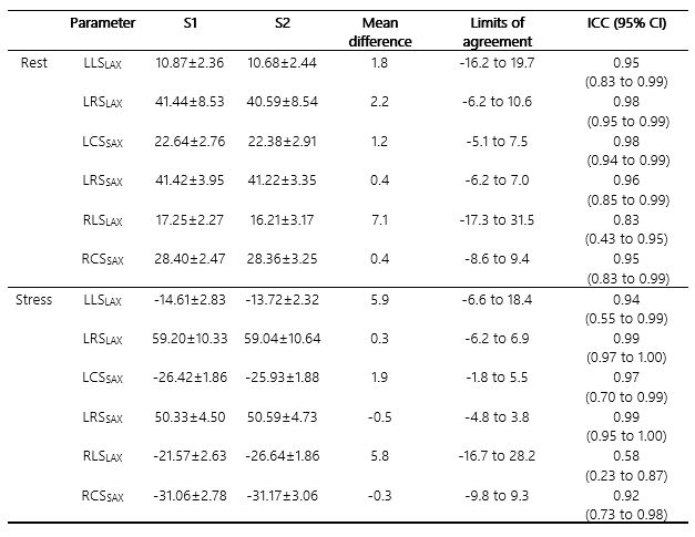
Statistical significance of the differences was assessed applying an unpaired Mann-Whitney U test, with p-values below 0.05 being considered significant. The intra-observer reproducibility was assessed using the intraclass correlation coefficient (ICC) and Bland-Altman analysis. Agreement was considered excellent for ICC > 0.74, good for ICC 0.6-0.74, fair for ICC 0.40-0.59, and poor for ICC < 0.40.
Results
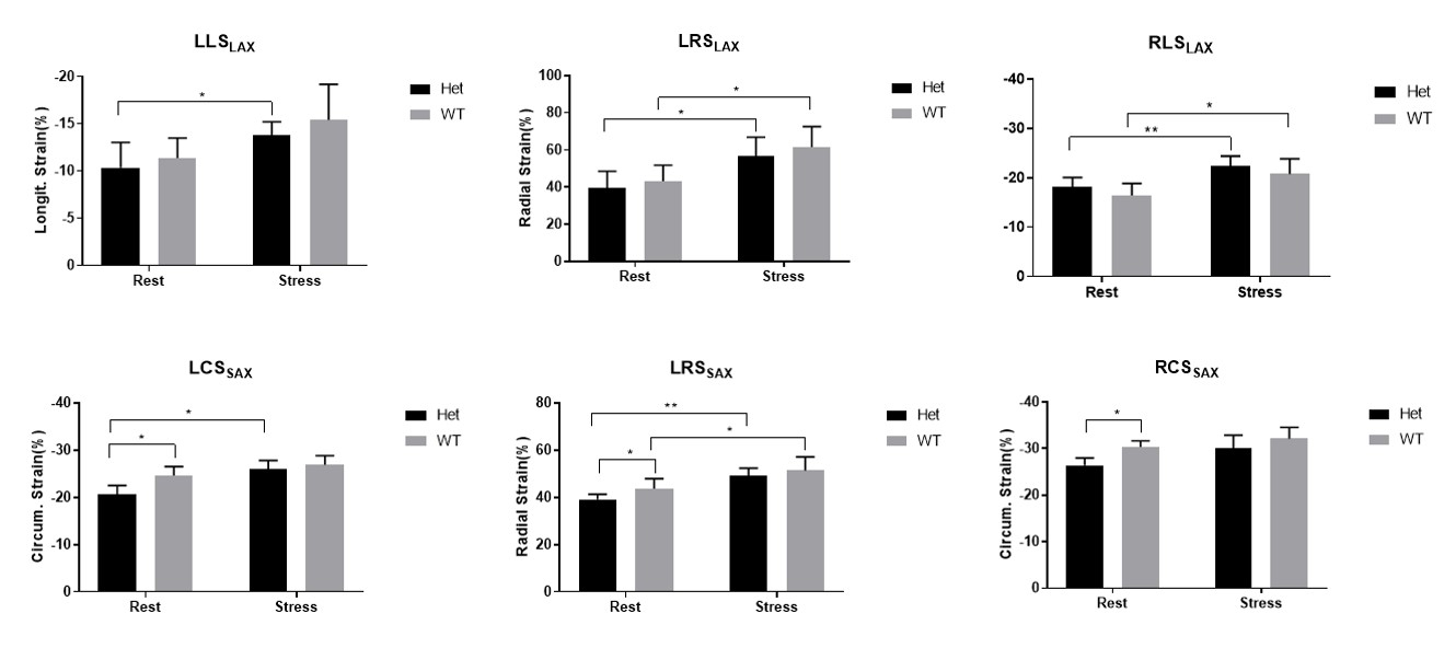
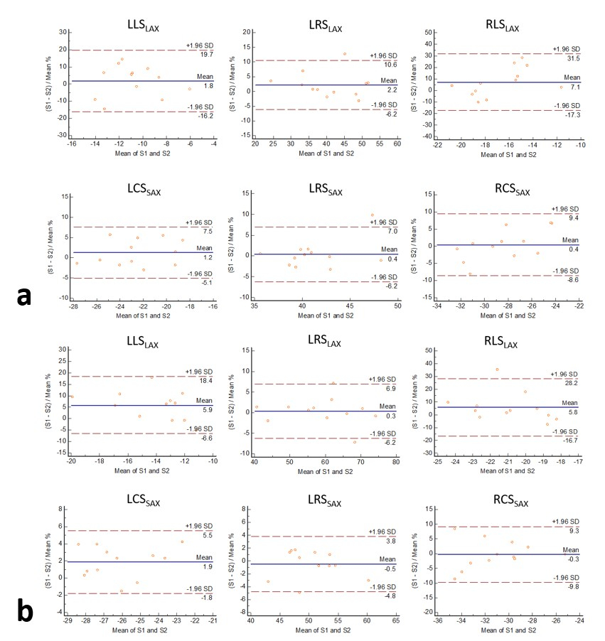
The acquisition protocol could be completed in all mice. Demographic characteristics, volumetric and functional parameters of study population were summarized in Table 1. A significantly decreased LV ejection fraction (EF), enlarged volume at end-diastole (EDV) and end-systole (ESV) were observed (p < 0.05). For the strain assessments at rest, a significantly higher circumferential strain and radial strain at short-axis could be observed in the WT group (p < 0.05), no difference was shown in long-axis. After dobutamine injection, myocardial strain increased in both groups (Fig.1).For the intra-observer reproducibility assessment, the RV longitudinal strain at stress showed an only fair reproducibility, while all other LV and RV strain assessments at rest and stress were excellent (Table 2). Bland-Altman plots demonstrate intra-observer reproducibility for left and right myocardial strain assessments (Fig. 2).
Conclusion
The analysis of cardiac myocardial strain derived from high resolution conventional cine images using CMR-FT technique provides a highly reproducible method for assessing myocardial performance in small animal models, expecially for LV strain analysis. For right ventricle, the circumferential strain at short-axis showed excellent reproducibility. However, the longitudinal strain at long-axis only showed fair reproducibility. The inter-observer and inter-study reproducibility will be addressed in the future.Acknowledgements
No acknowledgement found.References
1. Zerhouni EA, Parish DM, Rogers WJ, et al. Human heart: tagging with MR imaging--a method for noninvasive assessment of myocardial motion. Radiology. 1988 Oct;169(1):59-63.
2. Maret E, Todt T, Brudin L, et al. Functional measurements based on feature tracking of cine magnetic resonance images identify left ventricular segments with myocardial scar. Cardiovasc Ultrasound. 2009;7(1):53. doi:10.1186/1476-7120-7-53
3. Schuster A, Morton G, Hussain ST, et al. The intra-observer reproducibility of cardiovascular magnetic resonance myocardial feature tracking strain assessment is independent of field strength. European Journal of Radiology. 2013;82(2):296-301. doi:10.1016/j.ejrad.2012.11.012
4. Morton G, Schuster A, Jogiya R, et al. Inter-study reproducibility of cardiovascular magnetic resonance myocardial feature tracking. J Cardiovasc Magn Reson. 2012;14(1):43. doi:10.1186/1532-429X-14-43
5. Kempny A, Fernández-Jiménez R, Orwat S, et al. Quantification of biventricular myocardial function using cardiac magnetic resonance feature tracking, endocardial border delineation and echocardiographic speckle tracking in patients with repaired tetralogy of fallot and healthy controls. J Cardiovasc Magn Reson. 2012;14(1):32. doi:10.1186/1532-429X-14-32
6. Lapinskas T, Grune J, Zamani SM, et al. Cardiovascular magnetic resonance feature tracking in small animals – a preliminary study on reproducibility and sample size calculation. BMC Med Imaging. 2017;17(1):51. doi:10.1186/s12880-017-0223-7
7. Zuo Z, Subgang A, Abaei A, et al. Assessment of Longitudinal Reproducibility of Mice LV Function Parameters at 11.7 T Derived from Self-Gated CINE MRI. BioMed Research International. 2017;2017:1-10. doi:10.1155/2017/8392952
Figures