2157
Non-triggered, non-contrast lower extremity artery-weighted REACT : Impact of patient positioning for improving artery-vein contrast1Philips Japan, Tokyo, Japan, 2Department of Radiological Services, Tokyo Women's Medical University, Tokyo, Japan, 3Radiology, Eastern Chiba Medical Center, Chiba, Japan, 4Philips Healthcare, Tokyo, Japan
Synopsis
Artery-weighted REACT (REACT-Art), which is optimized the T2prep-time and TFE factor to improve the artery-vein contrast has been proposed, and it provide good artery-dominant contrast compared to conventional REACT. In this study, we focused on the impact of patient positioning to improve the artery-vein contrast with higher robustness, such as raising patient’s heels, putting patient’s heels down, compressing the patient’s legs. we have demonstrated that the patient-positioning like the patient legs to be compressed, and wearing compression stockings might achieve improved artery-vein contrast of REACT-Art with high stability and robustness in the lower extremities without use of contrast-enhancement and ECG-triggering.
Introduction
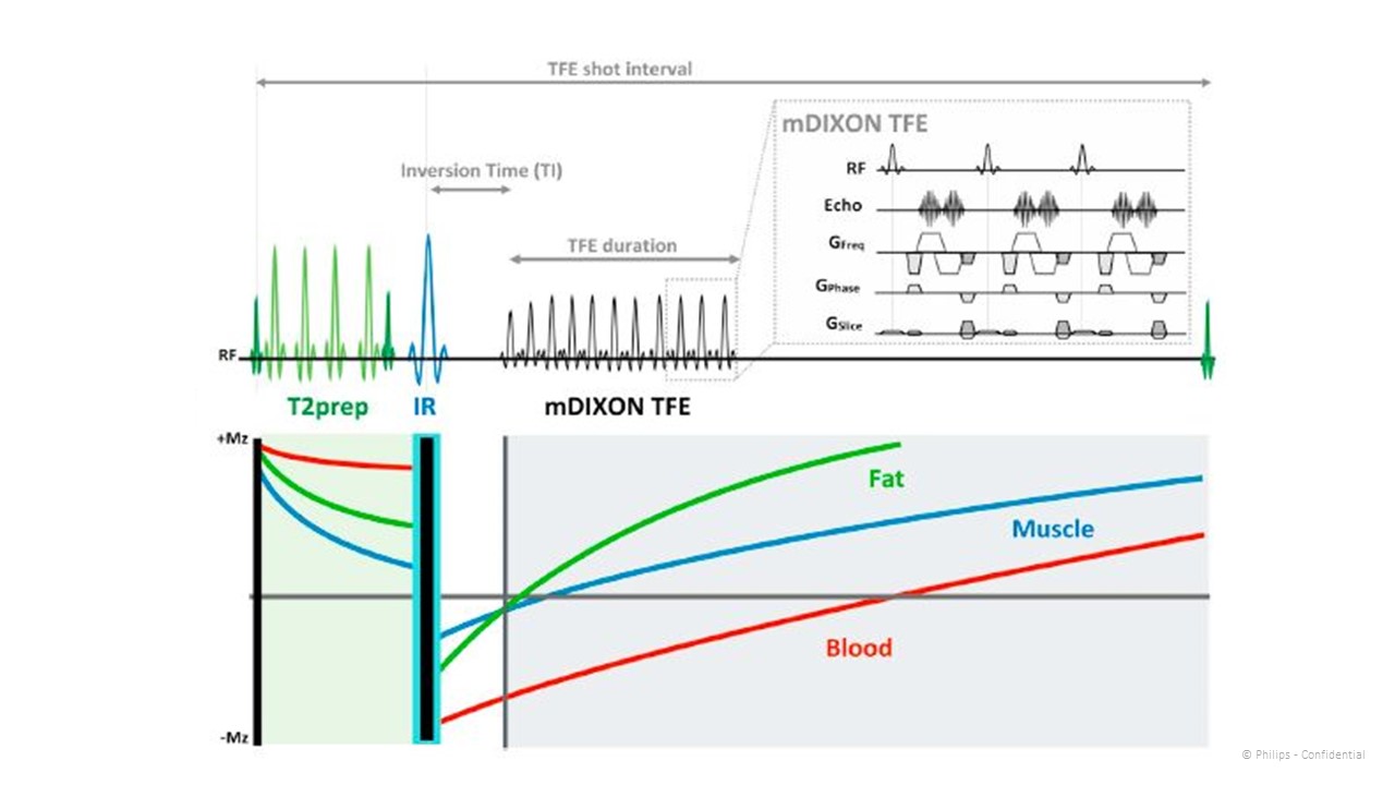
Relaxation-Enhanced MR Angiography without Contrast and Triggering (REACT) 1-3 derives high intravascular signal from T2 preparation pulse (T2prep) followed by STIR pulse and dual-echo gradient echo DIXON readout. A combination of T2prep, STIR pulses and DIXON emphasizes arterial and venous signal while effectively suppressing signal from surrounding tissues including muscle and fat. Conventional REACT sometimes cannot distinguish arteries and veins clearly. Recently, Artery-weighted REACT (REACT-Art), which was optimized the T2prep-time and TFE factor to improve the artery-vein contrast has been proposed, and it provided good artery-dominant contrast compared to conventional REACT in the lower extremities4. However, REACT-Art is still not enough to distinguish artery and vein consistently. One of the challenges of REACT-Art is to improve the artery-vein contrast with higher robustness. In this study, we focused on the impact of patient positioning, such as raising patient’s heels, putting patient’s heels down, compressing the patient’s legs. The purpose of this study is to investigate the impact of patient positioning and optimize the examination protocols including positioning to improve the artery-vein contrast in REACT-Art lower extremity MRA without ECG gating.Methods
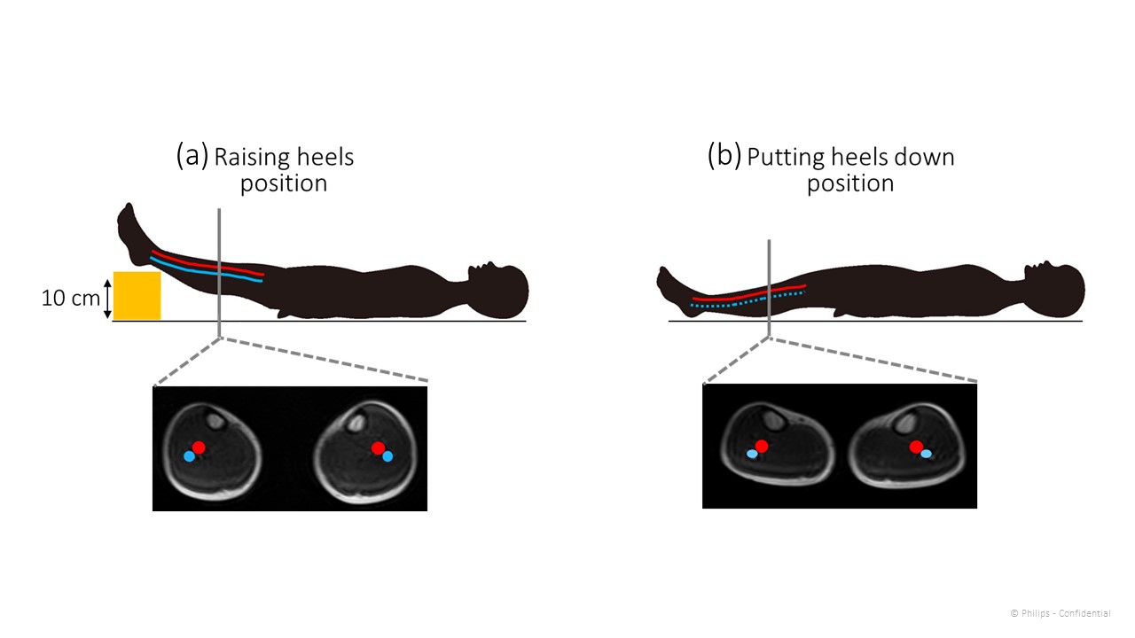
REACT-Art technique (Fig.1): REACT utilizes two pre-pulses and mDIXON-TFE, to exploit the long T1 and T2 relaxation times of unenhanced blood and to suppress background signals. A longer T2prep-time theoretically further suppresses venous signal because venous blood has shorter T2 value than arterial blood5-6.Subjects: REACT-Art in five healthy volunteers were examined on 3.0T MR scanner (Ingenia, Philips Healthcare) with below three situations (Fig. 2): 1. Raising the volunteer’s heels. 2. Putting the volunteer’s heels down. 3. Compressing the volunteer’s legs by wearing compression stockings. The volunteers obtained informed consent and approved by institutional review board.
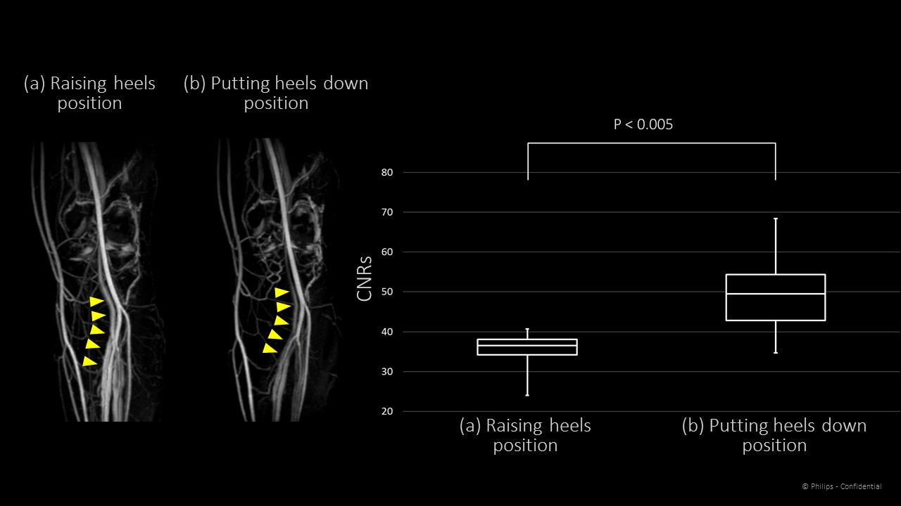
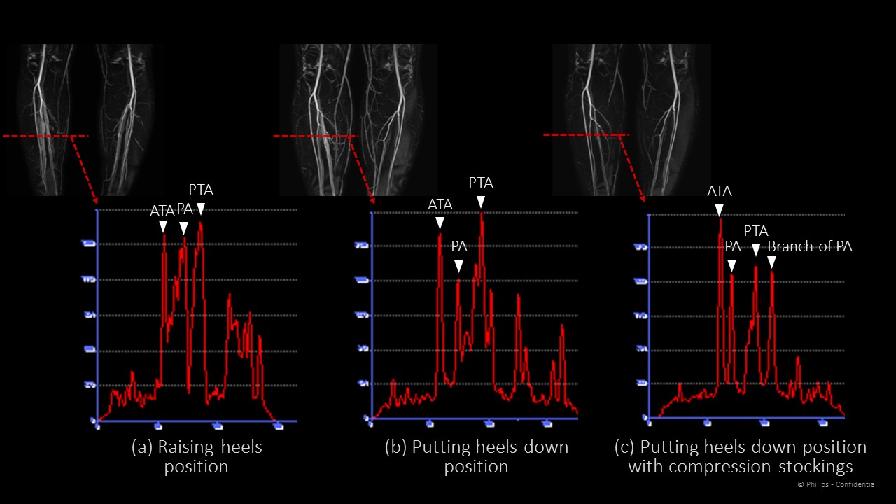
Evaluation: To compare the image qualities of REACT-Art with three situations, coronal maximum intensity projection (MIP) images were evaluated. Circular region of interests (ROIs) were placed on popliteal artery (PopA), popliteal vein (PopV) and background muscle. We evaluated contrast noise ratio (CNR) between signal intensity of PopA (SI PopA) and signal intensity of PopV (SI PopV) comparison of REACT-Art with raising heels and putting heels down situations. CNR was calculated as CNR = (SI PopA – SI PopV) / SD muscle. Here SI PopA and SI PopV is the respective mean signal intensity in the ROIs, and SD muscle is the standard deviation in the muscle ROI. The signal profile curve of REACT-Art with three situations in the anterior tibial artery (ATA), posterior tibial artery (PTA) and peroneal artery (PA) level evaluated.
Results
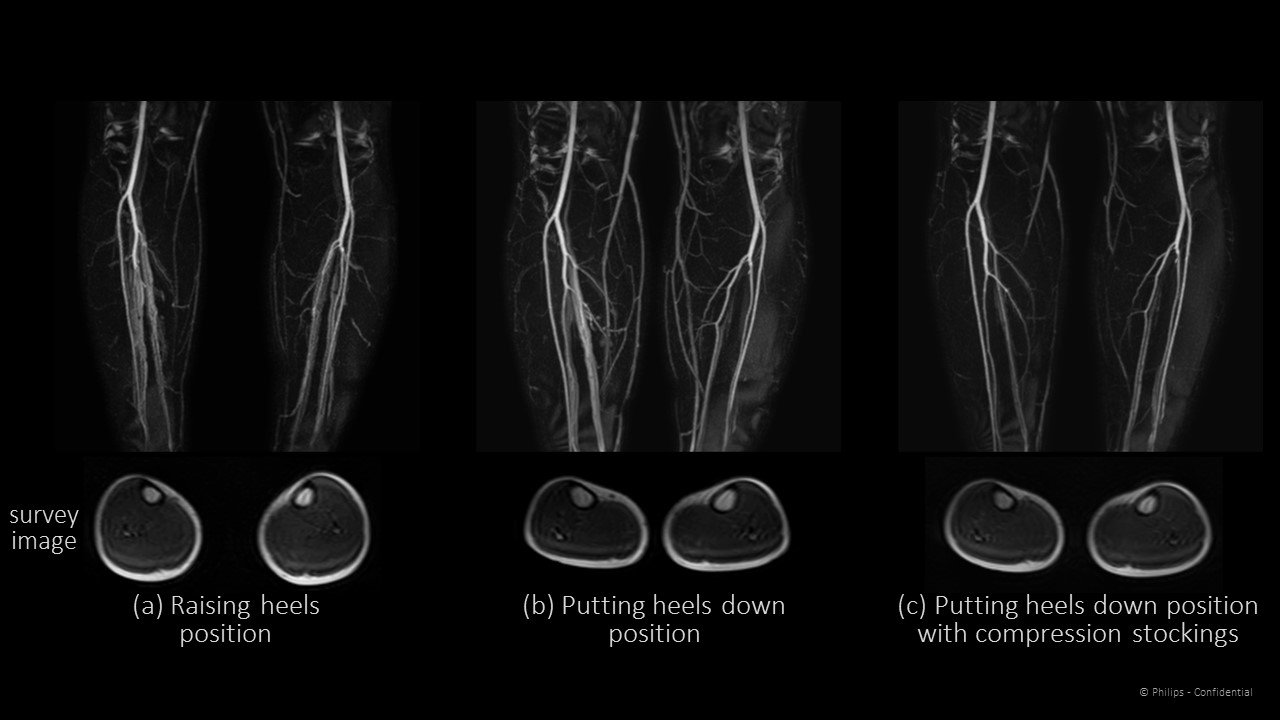
Fig. 3 shows the comparison of images and calculated CNRs among REACT-Art with raising the volunteer’s heels and putting the volunteer’s heels down. The CNR of REACT-Art with putting the volunteer’s heels down was higher than that of REACT-Art with raising the volunteer’s heels. There were significantly differences between two situations (p values < 0.005). Representative REACT-Art images with raising heels, putting heels down, and compressing legs by wearing compression stockings are shown in Fig. 4. REACT-Art with compressing the volunteer’s legs more suppressed vein signals compared to without compression. The signal profile curve of each vessel with compressing legs by wearing compression stockings was the sharpest compare to other situations.Conclusion
In this study, we have demonstrated that the patient-positioning like the patient legs to be compressed, and wearing compression stockings might achieve improved artery-vein contrast of REACT-Art with high stability and robustness in the lower extremities without use of contrast-enhancement and ECG-triggering.Acknowledgements
No acknowledgement found.References
1 Yoneyama M et al. Magn Reson Imaging. 2019 Aug 16;63:137-146.
2 Yoneyama M et al. In:Proc 24th Annual Meeting of ISMRM, Singapore 2016;2252.
3 Toyonari N et al. In:Proc 24th Annual Meeting of ISMRM, Singapore 2016;2684.
4 Hamano H et al. In:Proc 27th Annual Meeting of ISMRM, Montreal 2019;2070.
5 Brittain JH et al. Coronary Angiography with Magnetization-Prepared T2 Contrast. MRM. 1995; 33: 689-696.
6 Shea SM et al. Three-Dimensional True-FISP Imaging of the Coronary Arteries: Improved Contrast with T2-Preparation. JMRI. 2002; 15: 597–602
Figures