2149
Whole-Neck Arterial Evaluation with High Spatial Resolution using 3D Stack-of-Stars Quiescent Interval Slice-Selective (QISS) MRA1Radiology, NorthShore University HealthSystem, Evanston, IL, United States, 2The University of Chicago Pritzker School of Medicine, Chicago, IL, United States, 3Radiology, Northwestern University Feinberg School of Medicine, Chicago, IL, United States
Synopsis
We found that a prototype thin-slab stack-of-stars QISS sequence using a low-flip-angle FLASH readout can be used for whole-neck arterial evaluation in under 7 minutes. The technique minimizes flow-related saturation effects similarly to 2D QISS, while providing greater scan efficiency compared with 3D TOF and much improved image quality and through-plane spatial resolution compared with 2D TOF.
Introduction
Nonenhanced magnetic resonance angiography (MRA) of the entire neck can be obtained with 2D time-of-flight (TOF). However, the image quality is suboptimal due to limited through-plane spatial resolution, motion sensitivity, and flow saturation artifact. Recently, a 3D thin-slab stack-of-stars QISS (tsSOS-QISS) technique using a bSSFP readout demonstrated the potential for high-quality nonenhanced MRA of the renal and lower extremity arteries.1 We hypothesized that a prototype tsSOS-QISS pulse sequence using a low-flip-angle FLASH readout could be used to minimize flow-related saturation effects similarly to 2D QISS2, while providing greater scan efficiency compared with 3D TOF and much improved image quality and through-plane spatial resolution compared with 2D TOF.Methods
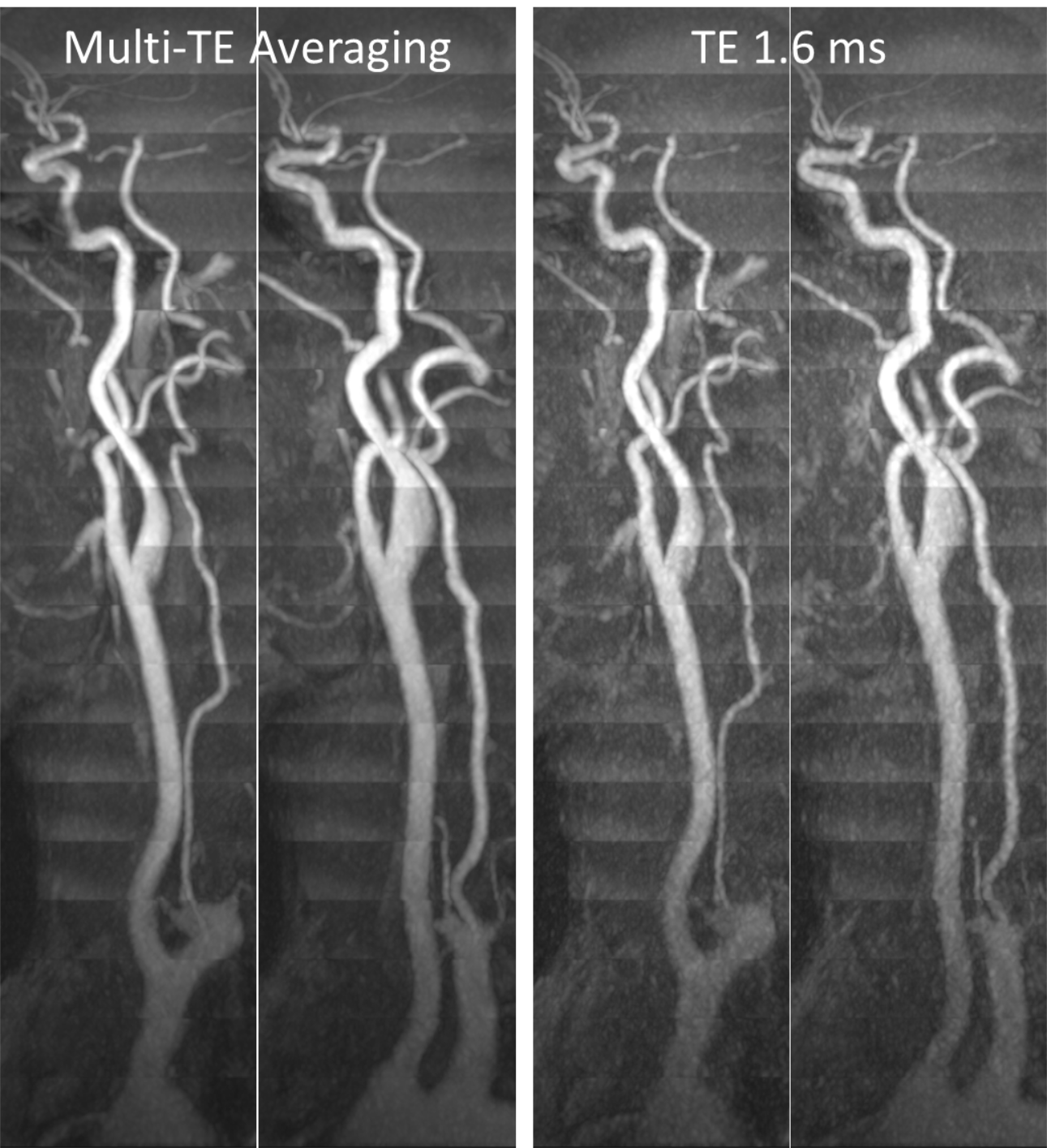
This study was approved by our institutional review board and all subjects provided written informed consent. Imaging was done on a 3 Tesla MRI system (MAGNETOM Skyrafit, Siemens Healthineers, Erlangen). Typical imaging parameters for 3D tsSOS-QISS were as follows: FLASH readout, TR of 9.9 ms, flip angle of 12-15°, 0.85 mm x 0.85 mm in-plane spatial resolution (reconstructed to 0.43 mm x 0.43 mm), 19-24 overlapping slabs with each slab providing 24-30 reconstructed 0.65 mm slices after two-fold interpolation in the slice direction (i.e. 12-15 acquired slices of 1.30 mm slice thickness), QISS TR/TI of 1500 ms/1040 ms with inversion-recovery based background and venous suppression, slab overlap of 23-25%, ramped radiofrequency pulses, 6/8th slice partial Fourier, receiver bandwidth of 590 Hz/pixel, scan times of ~6.6 min. A multi-echo readout with echo times (TEs) of 1.6 ms, 3.7ms, and 5.7 ms was used; the images formed from the three echoes could be averaged to improve signal-to-noise ratio. Comparisons of 3D tsSOS-QISS were made with 2D TOF, 2D QISS, as well as 3D TOF (TR/TE/flip 21ms/3.1ms/20°) acquired with equal spatial resolution, anatomical coverage, and scan time as 3D tsSOS-QISS.Signal measurements of 3D tsSOS-QISS were made in the carotid arteries and nearby muscle tissue. Arterial contrast-to-noise ratio (CNR) was estimated as (Sa-Sm)/σm, where Sa, Sm, and σm denote mean arterial signal, mean muscle signal, and standard deviation of muscle signal, respectively. CNR values from the composite image obtained by averaging the three echo time images were compared to those of the first TE of 1.6 ms.
Results
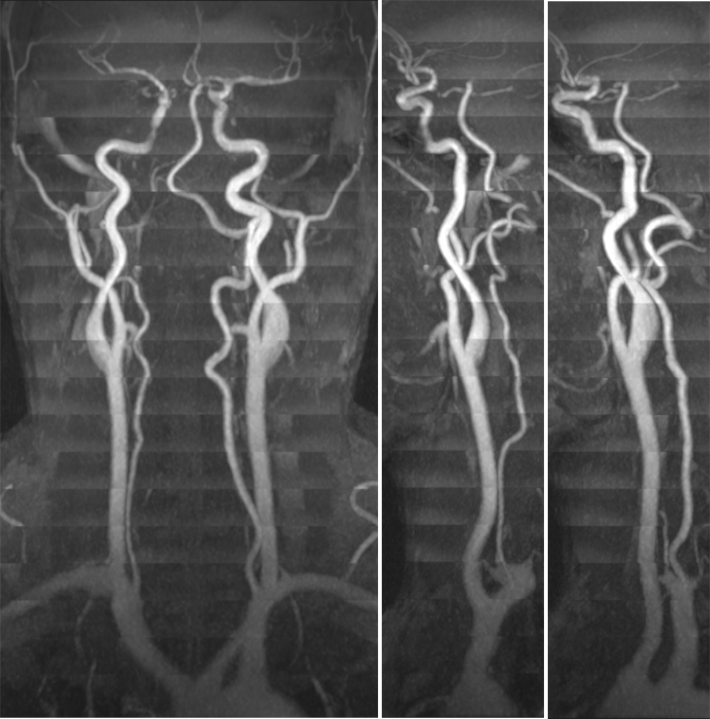
Figure 1 shows an example of 3D tsSOS-QISS MRA of the entire neck. It provided excellent image quality on both source images and multiplanar reconstructions, with excellent portrayal of the full lengths of the carotid and vertebral arteries. The averaging of images obtained at the three echo times increased CNR by 115% with respect to the first TE of 1.6 ms (Figure 2). Figure 3 shows tsSOS-QISS images in comparison to 2D QISS and 3D TOF. Compared to 2D TOF or 2D QISS, 3D tsSOS-QISS showed smoother vessel contours and was less sensitive to respiratory motion, with improved display of the carotid and vertebral artery origins. 3D tsSOS-QISS provided substantially better image quality than 3D TOF matched for spatial resolution, scan time, and anatomical coverage.Discussion
The results of this study show that 3D tsSOS-QISS is an efficient technique for whole-neck arterial evaluation without the use of contrast agents. In comparison with alternative approaches such as 2D TOF and 3D TOF, 3D tsSOS-QISS provides much smaller voxels and better image quality, respectively. Moreover, tsSOS-QISS allows targeted evaluation of the carotid bifurcation in <1 minute. The use of a low flip angle excitation (not possible with TOF due to loss of arterial-to-background contrast) reduces saturation of arterial signal. Future efforts will seek to validate the 3D tsSOS-QISS protocol in patients with suspected arterial pathology.Conclusion
We have demonstrated that 3D tsSOS-QISS enables near-isotropic high spatial resolution MRA for whole-neck arterial evaluation with diagnostic image quality in under 7 minutes. Moreover, the use of a multi-echo data acquisition with signal averaging of resulting images approximately doubled the contrast-to-noise ratio.Acknowledgements
Funding Source: NIH grant R01 EB027475References
1. Edelman RR, Aherne E, Leloudas N, Pang J, Koktzoglou I. Near-isotropic noncontrast MRA of the renal and peripheral arteries using a thin-slab stack-of-stars quiescent interval slice-selective acquisition. Magn Reson Med. 2019 Oct 21. doi: 10.1002/mrm.28032. [Epub ahead of print]
2. Koktzoglou I, Aherne EA, Walker MT, Meyer JR, Edelman RR. Ungated nonenhanced radial quiescent interval slice-selective (QISS) magnetic resonance angiography of the neck: Evaluation of image quality. J Magn Reson Imaging. 2019 May 11. doi:10.1002/jmri.26781. [Epub ahead of print]
Figures