2147
A comparative study of non-contrast enhanced 3D TRANCE and 3D Balanced FFE imaging of pulmonary artery with Time-SLIP
Yuan QU1, Yan WANG1, Yuli HUANG2, Li ZHU3, and Haiyang DONG3
1Radiology, People’s Hospital of Xinjiang Uygur Autonomous Region, Urumqi, China, 2MR, Philips Healthcare (Suzhou) Co., Ltd, SUZHOU, China, 3Radiology, Shanghai Chest Hospital, Shanghai JiaoTong University, Shanghai, China
1Radiology, People’s Hospital of Xinjiang Uygur Autonomous Region, Urumqi, China, 2MR, Philips Healthcare (Suzhou) Co., Ltd, SUZHOU, China, 3Radiology, Shanghai Chest Hospital, Shanghai JiaoTong University, Shanghai, China
Synopsis
The Time-Spatial Labeling Inversion Pulses (Time-SLIP) technique was used to perform non-contrast enhanced pulmonary artery imaging using 3D TRANCE and 3D Balanced FFE respectively, and the image quality obtained by the two imaging methods and the number of branches of pulmonary artery were compared specifically. Both 3D TRANCE and 3D Balanced FFE imaging methods with Time-SLIP can be used for pulmonary artery imaging. The image quality of pulmonary artery obtained by 3D TRANCE is generally better than 3D Balanced FFE.
INTRODUCTION
Pulmonary angiography mostly relies on techniques such as computed tomography angiography (CTA), digital subtraction angiography (DSA) and contrast-enhanced magnetic resonance angiography (CE-MRA).1,2 Alternatively, non-contrast enhanced MRA (NCE-MRA) sequences have been developed for characterizing structure of pulmonary artery.3 Time-spatial labeling inversion pulses (Time-SLIP) assist in morphological diagnosis via selectively observing the target vessels.4-6 In the present study, we investigated the performance of non-contrast enhanced 3D TRANCE without ECG triggered and 3D balanced FFE (3D-bFFE) using Time-SLIP on pulmonary artery imaging.METHODS
Study Sample: Twenty-three healthy subjects (10 males and 13 females, mean age 41.08 ± 13.67 years) were recruited for the study. This study was approved by local institutional review board and written consent form was obtained from each subject. MR Imaging: All pulmonary artery images were acquired on a 1.5 Tesla MR scanner (Achieva, Philips Healthcare) with an 8-channel abdominal coil. Each subject underwent NCE-MRA using 3D TRANCE and 3D-bFFE with Time-SLIP. 3D TRANCE sequence was performed with following parameters: repetition time 2000-3000ms, echo time 138ms, flip angle 90°, field of view 320x360mm2, slice number 60, slice thickness 3.0mm. 3D-bFFE sequence was performed with following parameters: repetition time 3.8ms, echo time 1.87ms, flip angle 65°, field of view 320x360mm2, slice number 60, slice thickness 3.0mm. Two kinds of spatial selective inversion labeling methods were used in this study: blood in superior and inferior vena cava, and blood in right ventricular and right atrium (Figure 1). The inversion time was set to 1200ms empirically. Data analysis: Signal-to-noise ratio (SNR) and contrast-to-noise ratio (CNR) in different regions of interest (ROIs) in the branches of pulmonary arteries were calculated using standard deviation in muscular tissue as noise. Additionally, the number of pulmonary artery and its branches was counted and the image quality was evaluated with 1 to 4 score (Figure 2). Image quality review was performed by two observers with >10 years’ experience in the department of radiology blinded to each other and the mean value of their outcome were used for further statistical analysis. Statistical analysis: The SNR and CNR were compared between 3D TRANCE and 3D-bFFE using a paired t-test. The Spearman`s rank sum test was performed to compare the subjective scores of image quality and the count of distinct branches of different orders. All statistical analyses were conducted with SPSS 25.0 (IBM Inc., USA).RESULTS
SNR and CNR of the pulmonary trunk, left and right pulmonary arteries, and their branches in Time-SLIP 3D TRANCE images were significantly better than those in Time-SLIP 3D-BFFE images (p<0.05). With 3D TRANCE, the count of order 3 and order 4 branches of left and right pulmonary arteries were statistically significantly more than 3D-bFFE (P<0.05). As for the results of image quality evaluation of pulmonary artery, 3D TRANCE was also significantly better than 3D-bFFE (P<0.05) (Table 1). When using 3D TRANCE, different positions of spatial selective inversion labelling band affected the visualization of pulmonary artery branches, such as the inferior branch of the left pulmonary artery and the inferior branch of the right pulmonary artery.DISCUSSION
TRANCE was a turbo spin echo sequence which acquires data with optimized suppression of background tissue and fat, resulting in better structure visualization of pulmonary artery. Compared to 3D-bFFE, TRANCE was insensitive to susceptibility and obtained better illustration of pulmonary artery and its branches. Thus, TRANCE with Time-SLIP could provide a distinct vascular structure in chest MRI for pulmonary artery (Figure 3).CONCLUSION
Time-spatial labeling inversion pulse (Time-SLIP) technique can be used for selective, non-enhanced magnetic resonance imaging of the pulmonary artery. Image quality and diagnostic value are greater with Time-SLIP in combination with the 3D TRANCE sequences based on TSE than in combination with 3D-bFFE. Different inversion labeling band positions have a larger impact on visualization of pulmonary artery branches in Time-SLIP with 3D TRANCE than with 3D-bFFE.Acknowledgements
No acknowledgement found.References
- Furlan A, Aghayev A, Chang CC, et al. Short-term mortality in acute pulmonary embolism: clot burden and signs of right heart dysfunction at CT pulmonary angiography. Radiology, 2012, 265(1): 283-293.
- Rashid H N Z, Lim A K, Lau K K. Use of Computed Tomography – Digital Subtraction Angiography in differentiating pulmonary thrombosis and pulmonary artery dissection in a large pulmonary artery aneurysm: [J]. Respiratory Medicine Case Reports, 2016, 18:24-26.
- Miyazaki M, Akahane M. Non-contrast enhanced MR angiography: Established techniques[J]. Journal of Magnetic Resonance Imaging, 2012, 35(1):1-19.
- Hamamoto K, Chiba E, Matsuura K, et al. Non–contrast-enhanced magnetic resonance angiography using time-spatial labeling inversion pulse technique for differentiation between pulmonary varix and arteriovenous malformation[J]. Radiology Case Reports, 2017: S1930043316303880.
- Kohei H, Katsuhiko M, Emiko C, et al. Feasibility of Non-contrast-enhanced MR Angiography Using the Time-SLIP Technique for the Assessment of Pulmonary Arteriovenous Malformation[J]. Magnetic Resonance in Medical Sciences, 2016, 15(3):253-265.
- Shonai T, Takahashi T, Ikeguchi H, et al. Improved arterial visibility using Short-Tau Inversion-Recovery (STIR) fat suppression in non-contrast-enhanced Time-Spatial Labeling Inversion Pulse (Time-SLIP) renal MR Angiography (MRA)[J]. Journal of Magnetic Resonance Imaging. 2009, Jun;29(6):1471-1477.
Figures
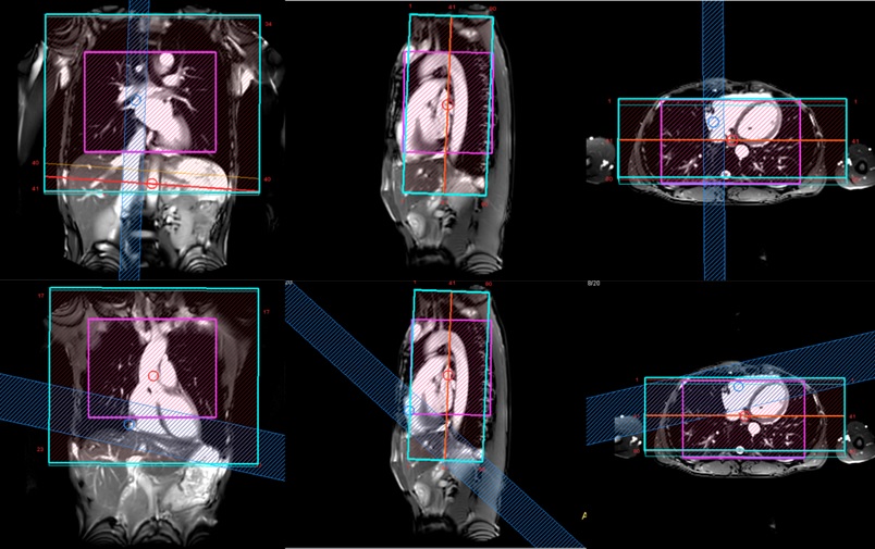
Figure 1. The placement of the Time-SLIP over
the superior vena cava (SV) and inferior vena cava (IV) (top row). The placement
of the Time-SLIP over the right atrium (RA) and right ventricle (RV) (bottom
row).

Figure 2. Subjective assessment criteria for
pulmonary image quality. The score of image (a) is 4; image (b) is 3; image (c)
is 2; image (d) is 1.
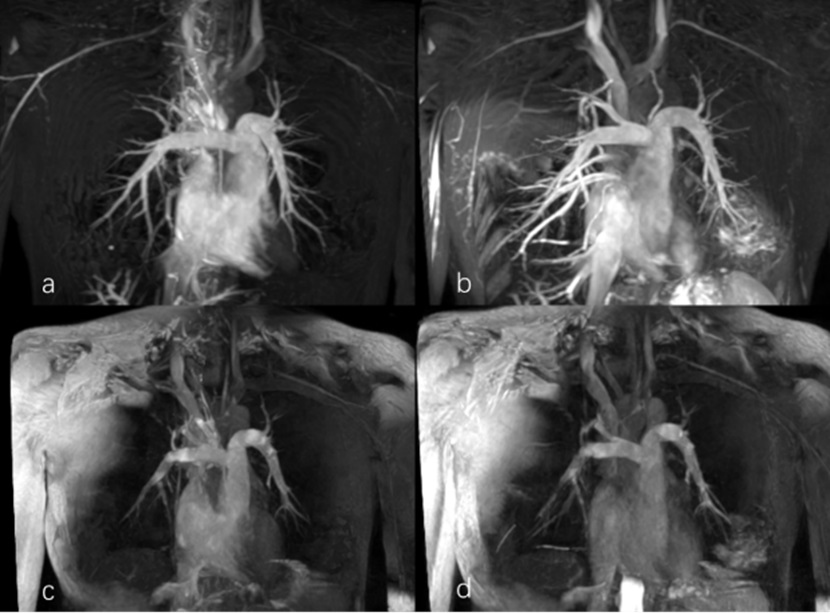
Figure 3. Pulmonary artery imaging. (a) Time-SLIP
3D TRANCE, the selective inversion labelling band covers the SV and IV. (b) Time-SLIP
3D TRANCE, the inversion band covers the RA, RV. (c) Time-SLIP 3D-bFFE, the inversion
band covers the SV and IV. (d) Time-SLIP 3D-bFFE, the inversion band to cover
the RA, RV. The 3D TRANCE images illustrate better structure of pulmonary
artery and its branches with optimized background tissue and fat suppression.
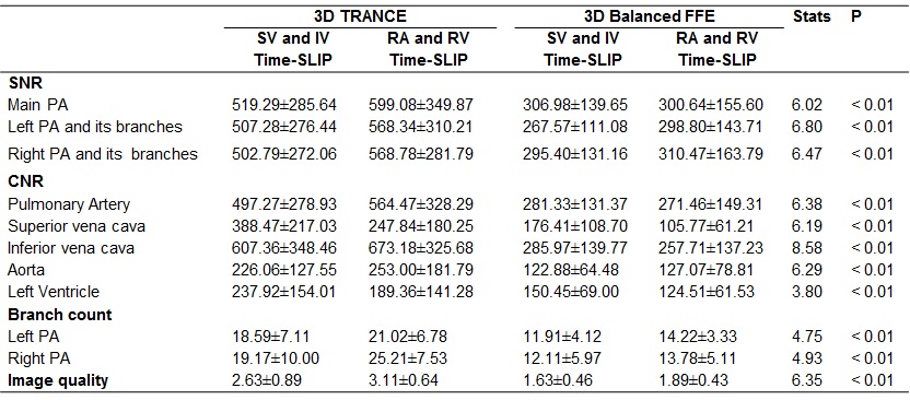
Table1. Statistical results of the
SNR and CNR, the number of branches, image quality for pulmonary artery (PA) images
between 3D TRANCE and 3D-bFFE.