2143
Non-contrast enhanced angiography and vessel wall imaging of coronary artery at 3T MR1Radiology, Fuwai hospital chinese academy of medical science, shenzhen, Shenzhen, China
Synopsis
Non-contrast enhanced MR angiography of coronary artery is a promising imaging method. In this study, the image quality of two non-contrast enhanced coronary MRA imaging methods were compared, included 3D-mDIXON and 3D-TFE-WHCA. 3D-mDIXON and 3D-TFE-WHCA showed good image quality with high SNR, and 3D-TFE-WHCA showed improved CNR and higher score in vascular display quality. According CTA data, T1-TFE-BB demonstrated the coronary vessel wall as medium high signal with regular margin and good contrast with the fat around the coronary artery. Combination of 3D-TFE-WHCA and T1-TFE-BB can be used as the imaging research method of coronary atherosclerosis.
Introduction
Non-contrast enhanced MR angiography of coronary artery is a promising imaging method without exposing the patient to radiation1-3. It is important to detect coronary artery stenosis and vessel wall for coronary MRA. The imaging methods of coronary artery vessel wall are still under development4. The purpose of this study is to investigate the imaging characteristics of 3D-mDIXON and 3D-TFE-WHCA and the high resolution vessel wall imaging, so as to propose the MR research method of coronary atherosclerosis.Objectives
To compare the image quality of 3D-mDIXON and 3D-TFE-WHCA for Non-contrast enhanced coronary artery MR angiography and to confirm the feasibility of T1-TFE with black-blood (T1-TFE-BB) as coronary vessel wall imaging.Materials and methods
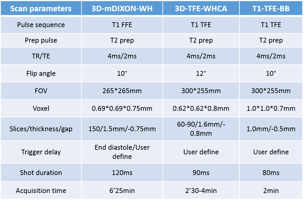
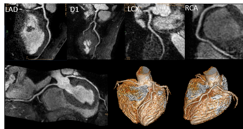
3D-mDIXON and 3D-TFE-WHCA Coronary MRA and T1-TFE-BB vessel wall imaging were performed on a 3T scanner (Ingenia, Philips Healthcare, the Netherlands) using body multichannel coil under breathing navigation. Imaging data were acquired in 29 patients (15 male, 14 female, mean age 53±10 years), excluded those with arrhythmias. The scanning parameters of 3D-mDIXON, 3D-TFE-WHCA and T1-TFE-BB were listed in table 1. The signal intensity of blood, fat and myocardium was measured with region of interest technique on the images of left main coronary bifurcation. Signal–to-noise ratio (SNR), contrast-to-noise ratio (CNR) of 3D-mDIXON and 3D-TFE-WHCA were compared. 8 patients underwent coronary CTA before or after MR examination within 2 months, and the results of coronary CTA were negative. According coronary CTA imaging, the vascular display quality of coronary MRA and T1-TFE-BB vessel wall imaging of 8 patients were scored (0-4 points) and compared.Results
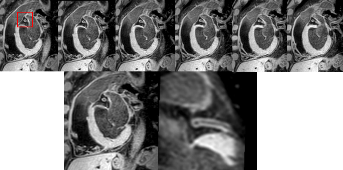
There was no statistical difference between 3D-mDIXON and 3D-TFE-WHCA in SNR (SNR: mDIXON=185.85±49.33 and WHCA=209.58±62.04, p=0.06). 3D-TFE-WHCA showed improved CNR compared to 3D-mDIXON (CNRblood-fat: mDIXON=147.73±46.15 and WHCA=118.98±54.54, p=0.03; CNRblood-myocardium: mDIXON=68.73±28.45 and WHCA=209.58±62.04, p=0.00). In 8 patients with negative coronary CTA results, 3D-TFE-WHCA showed higher score of vascular display quality in the middle or distal segment of coronary artery (RCAmiddle: mDIXON=2.5±1.2 and WHCA=3.1±0.7, p=0.01; PDA: mDIXON=2.7±0.9 and WHCA=3.0±0.6, p=0.01; LADdistal: mDIXON=2.8±0.9 and WHCA=3.0±0.7, p=0.03; LCXmiddle: mDIXON=2.6±0.9 and WHCA=2.9±0.9, p=0.03). The vessel wall of coronary artery was demonstrated as medium high signal with regular margin on T1-TFE-BB. In contrast, the fat around the coronary artery and the lumen were showed low signal. The scan range of T1-TFE-BB covered left main coronary and proximal anterior descending branch. There was no statistical difference between T1-TFE-BB and CTA in the quality score of vascular wall imaging (CTA =3.7±0.5, T1-TFE-BB =3.5±0.8, p=0.16 ).Conclusions
3D-mDIXON and 3D-TFE-WHCA can be used for non-contrast enhanced coronary MRA imaging. 3D-TFE-WHCA has higher image quality and better display effect for distal segments of coronary arteries compared with 3D-mDIXON. Combination of 3D-TFE-WHCA and T1-TFE-BB can be used as the imaging research method of coronary atherosclerosis.Key words
coronary magnetic resonance angiography, mDIXON, vessel wallAcknowledgements
I would like to express my gratitude to all the authors for their contrubutions for this article. In particular, I gratefully acknowledge the help of Prof. Yuan Xuchun for his valuable suggestions and critiques. My gratitued also extends to my lovely family.References
1.Marc Dweck, Valentina Puntmann, Alex Vesey, et al. MR imaging of coronary arteries and plaques. JACC: cardiovascular imaging, 2016,9(3):306-16.
2.Maryam Nezafat, Markus Hennlngsson, David Rlpley, et al. Coronary MR angiography at 3T: fat supperssion versus water-fat separation. Magn Reson Mater Phy, 2016,29:733-738.
3.Iyama Y, Nakaura T, Nagayama Y, et al. Single-Breath-Hold Whole-heart Unenhanced Coronary MRA Using Multi-shot Gradient Echo EPI at 3T: Comparison with Free-breathing Turbo-field-echo Coronary MRA on Healthy Volunteers [J]. Magnetic Resonance in Medical Sciences, 2018, 17(2):161-167.
4.Yibin Xie, Youngjin Kim, Jianing Pang, et al. Coronary atherosclerosis T1-weighted characterization with integrated anatomical reference. JACC: cardiovascular imaging, 2017,10(6):637-48.
Figures
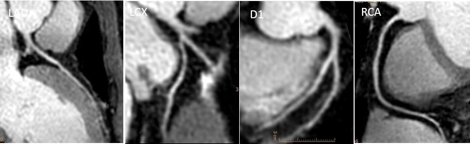
3D-TFE-WHCA MRA reconstruction images for coronary artery