2088
Pancreatic iron and cardiac iron and function in patients with thalassemia intermedia1MRI Unit, Fondazione G. Monasterio CNR-Regione Toscana, Pisa, Italy, 2"ARNAS" Civico, Di Cristina Benfratelli, Palermo, Italy, 3Ospedale "Sandro Pertini", Roma, Italy, 4Ospedale “SS. Annunziata” ASL Taranto, Taranto, Italy, 5Fondazione di Ricerca e Cura "Giovanni Paolo II", Campobasso, Italy, 6Azienda Ospedaliera "Garibaldi" Presidio Ospedaliero Nesima, Catania, Italy, 7Universitaria Careggi, Firenze, Italy, 8Ospedale San Martino di Oristano, Oristano, Italy
Synopsis
In patients with thalassemia intermedia (TI) pancreatic iron overload is frequent (58.1%), especially if regular transfusions are performed. Pancreatic T2* values were correlated with cardiac T2* values and a normal pancreas T2* showed negative predictive value of 100% for cardiac iron. Global pancreas T2* values were not correlated to biventricular volumes and ejection fraction or to myocardial fibrosis.
Introduction
In thalassemia major (TM) pancreatic iron was shown to be correlated to cardiac iron, biventricular function and myocardial fibrosis detected by cardiovascular magnetic resonance (CMR).1,2In the present multicenter study we explored the link between pancreatic iron and CMR parameters in a cohort of patients with thalassemia intermedia (TI).
Methods
We considered 155 TI patients (81 F, mean age 41.74±13.31 years) consecutively enrolled in the E-MIOT (Extension-Myocardial Iron Overload in Thalassemia) project.T2* measurements were performed over pancreatic head, body and tail and global value was the mean.3 Myocardial iron overload (MIO) was quantified using a T2* segmental approach.4 Biventricular function parameters were assessed by cine images.5 Late gadolinium enhancement (LGE) images were acquired to detect myocardial fibrosis.6
Results
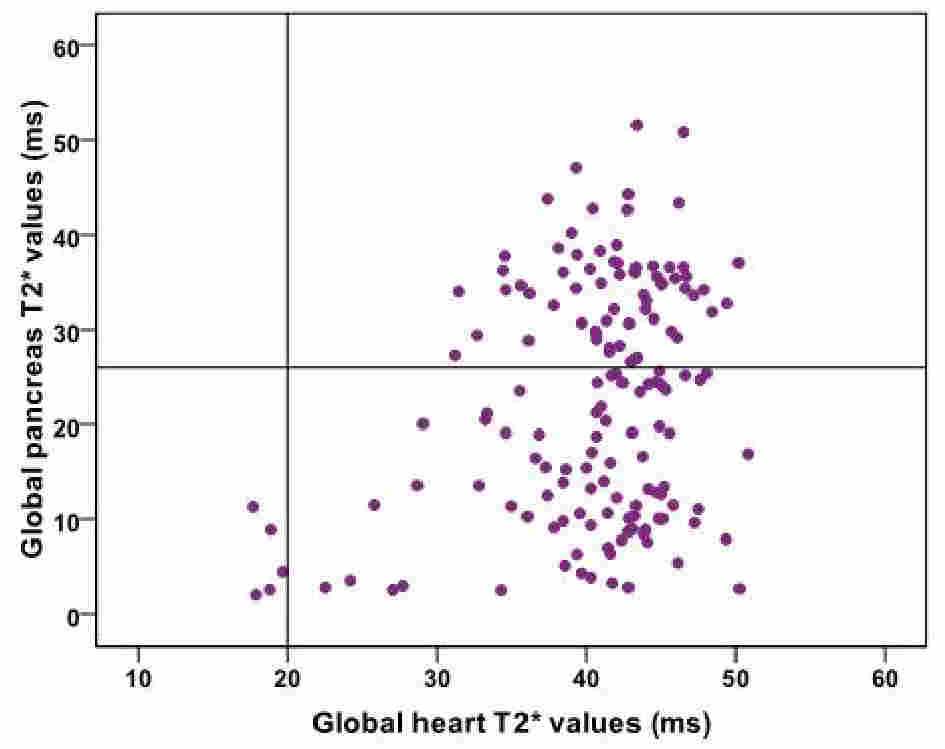
Global pancreas T2* values were not correlated to age or gender.Ninety patients (58.1%) showed a pathologic pancreas T2* value (<26 ms). One-hundred and five patients were transfusion dependent (TD) and they showed significantly lower global pancreas T2* values than non-TD patients (19.82±12.62 ms vs 28.52±10.81 ms; P<0.0001) and a significant higher frequency of pathologic global pancreas T2* value (66.7% vs 38.8%; P=0.001).
A significant correlation between pancreatic and cardiac T2* values was detected and (R=0.194; P=0.015) and all the 5 patients with MIO (global heart T2*<20 ms) had also pancreatic iron load (see Figure).
Global pancreas T2* values were not correlated to biventricular volumes and ejection fraction and no association was detected even in the subgroup of TD patients.
LGE sequences were acquired in 64 TI patients and 17 (26.6%) of them showed macroscopic myocardial fibrosis. Global pancreas T2* values were comparable in patients without and with fibrosis (24.37±11.65 ms vs 25.75±14.32 ms; P=0.574).
Conclusions
In TI patients a normal pancreas T2* showed negative predictive value of 100% for cardiac iron but pancreatic iron was not correlated to biventricular function and myocardial fibrosis.Acknowledgements
No acknowledgement found.References
1. Noetzli LJ, Papudesi J, Coates TD, Wood JC. Pancreatic iron loading predicts cardiac iron loading in thalassemia major. Blood 2009;114(19):4021-4026.
2. Meloni A, Restaino G, Missere M, et al. Pancreatic iron overload by T2* MRI in a large cohort of well treated thalassemia major patients: Can it tell us heart iron distribution and function? Am J Hematol 2015;90(9):E189-190.
3. Meloni A, De Marchi D, Positano V, et al. Accurate estimate of pancreatic T2* values: how to deal with fat infiltration. Abdom Imaging 2015;40(8):3129-3136.
4. Meloni A, Positano V, Pepe A, et al. Preferential patterns of myocardial iron overload by multislice multiecho T*2 CMR in thalassemia major patients. Magn Reson Med 2010;64(1):211-219.
5. Aquaro GD, Camastra G, Monti L, et al. Reference values of cardiac volumes, dimensions, and new functional parameters by MR: A multicenter, multivendor study. J Magn Reson Imaging 2016;45(4):1055-1067.
6. Pepe A, Positano V, Capra M, et al. Myocardial scarring by delayed enhancement cardiovascular magnetic resonance in thalassaemia major. Heart 2009;95:1688-1693.
Figures