2071
Cardiac Remodeling After Military Endurance Training: A Preliminary Study of Cardiac MRI Cine
Hongqin Liang1, Liqiang Zhu2, Bing Ji1, Qing Li3, Xiaoyue Zhou4, and Jian Wang1
1Southwest Hospital, Army Medical University, Chongqing, China, 2The Central Theater Command Air Force Hospital of PLA, Datong, China, 3Siemens Healthineers Ltd., Shanghai, China, 4Siemens Healthcare Ltd, Shanghai, China
1Southwest Hospital, Army Medical University, Chongqing, China, 2The Central Theater Command Air Force Hospital of PLA, Datong, China, 3Siemens Healthineers Ltd., Shanghai, China, 4Siemens Healthcare Ltd, Shanghai, China
Synopsis
Long-term military training leads to the remodeling of the heart and may cause exercise-induced myocardial injury (EIMI). The mechanism of heart remodeling is studied by using cardiac cine MRI to estimate left ventricular cardiac function and global stress. This study found that prolonged and intensive training lasting 4 years leads to cardiac morphological adaptations. Contrary to conventional thinking, left ventricular function changes happens earlier than the mechanical direction.
Introduction
High intensity and longtime training may lead to the remodeling of the heart in military personnel[1-3]. On the macro level, it is the enlargement of the morphological ventricle occurring that contributes to the remodeling. However, the mechanism of cardiac morphology changes is unclear. We hypothesized that cardiac remodeling occurs in military cadets who experienced training intensity and is caused by changes in the mechanical direction which precedes cardiac function.To explore the heart remodeling mechanism, cardiac cine MRI was used to quantify cardiac function and myocardial strain. The results between physically trained military personnel and senior students receiving no training were compared.Methods
The study comprised 66 volunteer subjects of which 43 were college senior students who received four years strength and endurance training of a certain intensity (5km run、Horizontal bar training 30 times per minute, push-ups 50 times per minute, sit-ups 50 times per minute ) in Chongqing, China, The control group includes 23 volunteers having no physical training (age = 21±2 years; 16 male and 7 female). MRI scans were performed on all subjects using a 3T MAGNETOM Trio a Tim system MR scanner(Siemens Healthcare, Erlangen, Germany). Subject information was collected and included data from the heart short axis, 2 cavity axes, and the 4 chamber heart film. The imaging protocols were including 3 short-axis slices (basal, middle, and apical LV) and a four-chamber slice using an electrocardiogram (ECG)-gated, breath-hold balanced steady-state free-precession (bSSFP) sequence with the following parameters: slice thickness = 6 mm, time of echo = 1.7ms, field of view (FOV) = 325 x 400 mm², matrix = 256 x 256, gap = 1.5 mm. The commercial software cvi42 (Circle Cardiovascular Imaging Inc., Calgary, Alberta, Canada) was utilized for post-processing using the 3D short module and 3D tissue tracking module. The left ventricular cardiac function and overall stress were measured, including global radial strain (GRS), global circumferential strain (GCS) and global longitudinal strain (GLS). The SPSS19.0 (IBM Corp., Armonk/NY, USA) was used for data analysis. The differences of each value were compared by independent sample t tests and P<0.05 was considered statistically significant.Results
Fig. 1 shows the comparisons of the cardiac function parameters between two groups. The difference of the cardiac function parameters between two groups which is statistically significant. The cardiac function parameters of experimental group were significantly higher than those of the control group except ejection fraction ( EF)% and heart rate(HR). Fig. 2 shows the difference of global strain parameters between two groups. The GRS peak strain and GLS peak diastolic strain rate of the experimental group are significantly difference ,and they are lower than those of the control group.Discussion
Military physical training increases the risk of EIMI[4-7]. However, this effect has not yet been previously studied using MRI cine, which acquires cardiac function information and strain parameters. Results of the present study showed that the cardiac function parameters of military personnel were higher than those of senior students, except for (EF)% and HR. No statistical difference in all strain parameters except for GRS peak strain and GLS diastolic strain rate, indicating that the change of strain direction was later than the change of function. The decrease of strain value in the longitudinal and radial direction in the experimental group indicated the decrease of myocardial elasticity, which may be the reason for the increase of cardiac morphology and the decrease of ejection fraction and heart rate. and which contributes to the understanding, diagnosis of heart Remodeling and EIMI.Conclusions
Four years of prolonged and intensive endurance training leads to cardiac morphological adaptations in military young subjects within this study. Contrary to our hypothesis, Initial results showed that left ventricular function changes earlier than the mechanical direction, at the same time, some indexes of mechanical changes have great influence on cardiac function parameters.Acknowledgements
No acknowledgement found.References
1. La Gerche A, Claessen G, Dymarkowski S, et al. Exercise-induced right ventricular dysfunction is associated with ventricular arrhythmias in endurance athletes. Eur Heart J, 2015, 36(30):1998-2010. 2. Eijsvogels TMH, Oxborough DL, O'Hanlon R, et al. Global and regional cardiac function in lifelong endurance athletes with and without myocardial fibrosis. European journal of sport science, 2017, 17(10):1297-303. 3. Gati S, Sharma S, Pennell D. The Role of Cardiovascular Magnetic Resonance Imaging in the Assessment of Highly Trained Athletes. JACC Cardiovasc Imaging, 2018, 11(2):247-59. 4. Bohm P, Schneider G, Linneweber L, et al. Right and Left Ventricular Function and Mass in Male Elite Master Athletes: A Controlled Contrast-Enhanced Cardiovascular Magnetic Resonance Study. Circulation, 2016, 133(20):1927-35. 5. Levine B D. Can intensive exercise harm the heart? The benefits of competitive endurance training for cardiovascular structure and function. Circulation, 2014, 130(12): 987-991. 6. Scally C, Rudd A, Mezincescu A, et al. Persistent Long-Term Structural, Functional, and Metabolic Changes After Stress-Induced (Takotsubo) Cardiomyopathy. Circulation, 2018, 137(10):1039-48. 7. Claus P, Omar AMS, Pedrizzetti G, et al. Tissue Tracking Technology for Assessing Cardiac Mechanics: Principles, Normal Values, and Clinical Applications. JACC Cardiovasc Imaging, 2015, 8(12):1444-60.Figures

Fig. 1 shows the
difference of the cardiac function parameters between two groups which is
statistically significant.
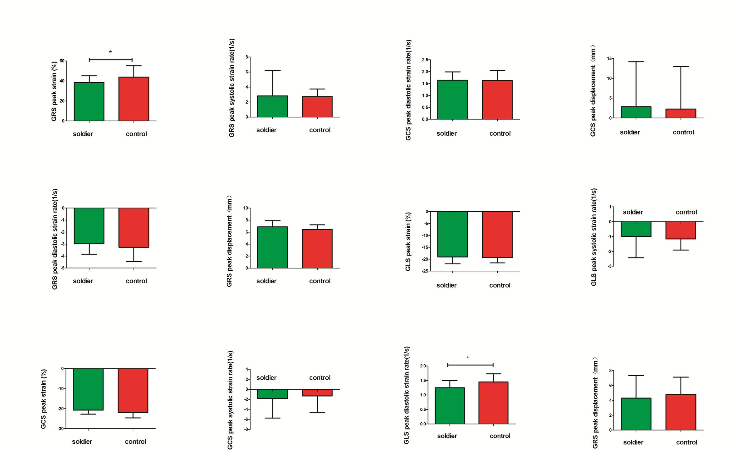
Fig. 2 shows the
difference of global strain parameters between two groups ,and which was
statistically significant between the GRS peak strain and GLS peak diastolic
strain rate only.